Published online Sep 28, 2006. doi: 10.3748/wjg.v12.i36.5902
Revised: August 16, 2006
Accepted: August 23, 2006
Published online: September 28, 2006
A 55-year old man presented with acute sigmoid volvulus. The distal level of obstruction was above the level which could be reached by the rigid sigmoidoscope to allow decompression, and so a flatus tube was "lassoed" onto the side of a flexible endoscope which allowed accurate placement under direct vision. This technique allows accurate placement of catheters, feeding tubes and other devices endoscopically, which cannot be placed through the instrument channel of the endoscope.
- Citation: Gatenby PAC, Elton C. Endoscopic placement of flatus tube using "lasso" technique with snare wire. World J Gastroenterol 2006; 12(36): 5902-5903
- URL: https://www.wjgnet.com/1007-9327/full/v12/i36/5902.htm
- DOI: https://dx.doi.org/10.3748/wjg.v12.i36.5902
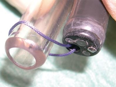
Acute sigmoid volvulus is frequently encountered in clinical practice. The distal level of obstruction can be reached by the rigid sigmoidoscope to allow decompression. A flatus tube (Figure 1) can be “lassoed” onto the side of a flexible endoscope which allows accurate placement under direct vision. This technique allows accurate placement of catheters, feeding tubes and other devices endoscopically, which cannot be placed through the instrument channel of the endoscope.
A 55-years old male presented acutely to the surgical on-call team at our institution with a 2-d history of absolute constipation and abdominal distension followed by vomiting. He had a previous history of laryngotomy for carcinoma and hypertension.
Clinical examination revealed abdominal distension, obstructive bowel sounds and an empty rectum. Plain abdominal radiology demonstrated features of distal large bowel obstruction and computed tomography revealed colonic distension to the level of the sigmoid colon, indicating a sigmoid volvulus and decompression was planned.
Under intravenous sedation with propofol, the colonoscope was passed into the sigmoid colon and the proximal colon was decompressed, but on removal of the endoscope, the sigmoid colon re-volved above the proximal extent of the rigid sigmoidoscope.
We employed a novel method for placement of the flatus tube under direct vision by ‘lassoing’ an endoscopic snare wire (passed through the colonoscope) onto the end of the flatus tube. The free end of the flatus tube was clamped to prevent escape of insufflated air during placement. The colonoscope (with ‘lassoed’ flatus tube) was renegotiated into the sigmoid colon and once satisfactory positioning was achieved under direct vision, the snare was loosened and slipped over the distal end of the flatus tube, which was left in place after the scope was withdrawn. The flatus tube was unclamped and left in-situ with a drainage bag attached for 48 h. The patient was allowed full diet after the procedure and discharged on the third day.
Acute sigmoid volvulus is frequently encountered in clinical practice. However, no previous report has described use of an endoscopic snare to guide the placement of either a hollow tube (to allow drainage or installation of substances) or a measuring catheter under direct vision. We employed a novel method for placement of the flatus tube under direct vision by ‘lassoing’ an endoscopic snare wire (passed through the colonoscope) onto the end of the flatus tube. The patient was allowed full diet after the procedure and discharged on the third day. It is suggested that this technique can achieve rather good results in the treatment of acute sigmoid volvulus. This technique can also be employed for upper alimentary procedures such as the accurate placement of feeding tubes or pH catheters.
S- Editor Wang GP L- Editor Wang XL E- Editor Bai SH


