Published online Nov 7, 2005. doi: 10.3748/wjg.v11.i41.6518
Revised: March 21, 2005
Accepted: March 24, 2005
Published online: November 7, 2005
AIM: To investigate the effect of H pylori eradication on atrophic gastritis and intestinal metaplasia (IM).
METHODS: Two hundred and fifty-nine patients with atrophic gastritis in the antrum were included in the study, 154 patients were selected for H pylori eradication therapy and the remaining 105 patients served as untreated group. Gastroscopy and biopsies were performed both at the beginning and at the end of a 3-year follow-up study. Gastritis was graded according to the updated Sydney system.
RESULTS: One hundred and seventy-nine patients completed the follow-up, 92 of them received H pylori eradication therapy and the remaining 87 H pylori-infected patients were in the untreated group. Chronic gastritis, active gastritis and the grade of atrophy significantly decreased in H pylori eradication group (P<0.01). However, the grade of IM increased in H pylori -infected group (P<0.05).
CONCLUSION: H pylori eradication may improve gastric mucosal inflammation, atrophy and prevent the progression of IM.
-
Citation: Lu B, Chen MT, Fan YH, Liu Y, Meng LN. Effects of
Helicobacter pylori eradication on atrophic gastritis and intestinal metaplasia: A 3-year follow-up study. World J Gastroenterol 2005; 11(41): 6518-6520 - URL: https://www.wjgnet.com/1007-9327/full/v11/i41/6518.htm
- DOI: https://dx.doi.org/10.3748/wjg.v11.i41.6518
H pylori-infected gastric mucosa evolves through stages of chronic gastritis, glandular atrophy (GA) and intestinal metaplasia (IM) which is a main cause of intestinal type of gastric adenocarcinoma. H pylori infection induces gastric epicyte apoptosis. The index of apoptosis and proliferation decrease after H pylori eradication[1,2]. H pylori eradication can prevent gastric cancer. Whether the precancerous lesions are reversed or terminated after H pylori eradication is crucial to evaluate this therapy for a preventive purpose. To investigate the histological changes in the gastric mucosa, we followed up chronic atrophic gastritis patients who received H pylori eradication therapy for 3 years.
A total of 259 H pylori-infected patients, who were diagnosed as having antral atrophic gastritis (without organic gastropathy, e.g., ulcer, tumor, etc.) by gastroscopy and biopsy in our hospital during 1997 to 1999, were followed up in our study; 154 of them received H pylori eradication therapy and the remaining 105 did not receive H pylori eradication therapy. Patients who failed to respond to H pylori eradication therapy and had uncertain H pylori infection were excluded. Patients who did not return for re-evaluation or became H pylori negative without receiving H pylori eradication therapy were also excluded. A total of 179 patients including 82 men and 97 women aged 30-74 years (92 in the H pylori eradicated group and 87 in the control group) completed the 3-year follow-up study.
All the subjects underwent gastroscopy when they entered the study and fulfilled the 3-year follow-up; some patients underwent gastroscopy every year. During gastroscopy, 3 antral biopsy specimens (from the greater and lesser curvatures 2-3 cm from the pylorus) were taken. One antral biopsy specimen was submitted to rapid urease test (RUT) and the other two were cut into sections for histological examination.
The patients were confirmed to have H pylori infection only when both RUT and histological stain (modified Giemsa stain) produced positive results. Otherwise they were considered as non-H pylori-infected healthy volunteers. Uncertain cases that were positive in one test and negative in the other test should be excluded. After they were treated for more than 4 wk, H pylori status was determined by 14C-urea breath test.
Biopsy specimens were stained with H&E. A single pathologist, who was unaware of the conditions of the patients and assigned treatment of biopsies, performed the histological assessment. The gastritis was graded using the visual analog scales of the updated Sydney system[3] as none (0), mild (1), moderate (2), or marked (3). Histological grading of the same subject before and 3 years after the therapy was compared and classified as improvement, no improvement, or exacerbation.
To eradicate H pylori infection, the patients received omeprazole (20 mg) or lansoprazole (30 mg), clarithromycin (500 mg), amoxicillin (1 g) or furazolidone (0.1 g), twice daily for 1 wk. During the whole follow-up period, two group patients received expectant treatment.
The results of histology score were expressed as mean .
The differences between the two groups before treatment with respect to histological grades were calculated using Mann-Whitney U test. Changes in histological grading in H pylori eradication and control groups before and 3 years after the treatment were compared by Stuart-Maxwell test. Differences in the proportions of histological grading with improvement, no improvement and exacerbation between two groups were made using the χ2 test. P < 0.05 was considered statistically significant (two-sided).
The H pylori eradicated group and control group were comparable in age and sex ratio (P>0.05). At entry into the study, the percentage of mild and moderate GA was 75% in H pylori-eradiated group and 25% in control group. The percentage of mild, moderate and marked GA was 77%, 21.8% and 1.2%, respectively. IM was 51% in H pylori-eradicated group and 58.6% in control group.
Active (AI) and chronic inflammation (CI) were found in both groups. Overall, the pretreatment histological scores were comparable in two treatment groups (Table 1).
| H pylori-eradicated group (n = 92) | Control group (n = 87) | |
| Chronic inflammation | 1.95 ± 0.40 | 1.97 ± 0.32 |
| Active inflammation | 1.84 ± 0.41 | 1.90 ± 0.40 |
| Glandular atrophy | 1.25 ± 0.44 | 1.24 ± 0.46 |
| Intestinal metaplasia | 0.64 ± 0.76 | 0.63 ± 0.57 |
Histological scores in paired samples before and after 3-year treatment in two groups are shown in Table 2.
| H pylori-eradicated group (n = 92) | Control group (n = 87) | |||
| Baseline | 3-yr | Baseline | 3-yr | |
| Chronic inflammation | 1.95 ± 0.40 | 1.56 ± 0.53b | 1.97 ± 0.32 | 1.91 ± 0.47 |
| Active inflammation | 1.84 ± 0.41 | 0.39 ± 0.19d | 1.90 ± 0.40 | 1.78 ± 0.44 |
| Glandular atrophy | 1.25 ± 0.44 | 0.97 ± 0.83b | 1.24 ± 0.46 | 1.19 ± 0.82 |
| Intestinal metaplasia | 0.64 ± 0.76 | 0.73 ± 0.77 | 0.63 ± 0.57 | 0.83 ± 0.87a |
After cure of infection, scoring of CI, AI and GA decreased significantly (P < 0.01) and that of IM increased slightly with no significant difference (P > 0.05). In control group, scoring of IM increased significantly (P < 0.05), but that of other pathologic changes had no statistical significance (P > 0.05).
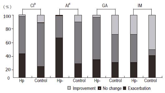
The histological changes in paired samples before and after 3-year treatment in H pylori-eradicated group and control group are shown in Figure 1. CI and AI were improved by 38.04% and 63.04% in H pylori-eradicated group, but only by 17.24% and 21.84% in the control group (P < 0.01). The GA was improved by 28.26% in the treatment group and 14.94% in the control group. GA was exacerbated by 4.35% in treatment group and 4.6% in control group (P > 0.05). IM was improved by 23.91% and exacerbated by 31.52% in H pylori-eradicated group, and by 20.69% and 33.33% in the control group (P > 0.05).
H pylori is one of the causative factors for gastric cancer. Its eradication may play an important role in the prevention of gastric cancer. GA and IM are pre-cancerous lesions, but data on the effects of anti-H pylori therapy on GA and IM are conflicting. Vander Hulst et al[4] reported that the severity of GA and IM does not change after treatment. Forbes et al[5]showed that no changes in GA and IM are to be found after H pylori eradication treatment.
Sung et al[6] showed that IM in the gastric antrum is slightly improved in H pylori-eradicated patients and GA in the corpus is significantly improved in H pylori- infected patients. Ohkusa et al[7]demonstrated that 89% of GA and 61% of IM are improved. Ito et al[8] have reported a marked decrease in the average grade of IM and GA after treatment. Correa et al[9]reported the proportions of GA and IM are increased significantly in H pylori-eradicated patients. Ruiz et al[10]showed that GA in the antrum improves significantly after H pylori eradication, but no significant change is observed by standard visual scoring method.
In our study, after a 3-year follow-up the scoring of GA decreased significantly in H pylori-eradicated group, while no significant change was found in control group (P < 0.01). The proportion of change in GA was higher in H pylori-eradicated group than in control group (28.26% vs 14.94%), but it was not statistically significant (P > 0.05). Though the GA grade in our study was mild and moderate, the results showed that eradication of H pylori was beneficial to some patients with GA in the antrum. Compared with GA, IM had no significant change after H pylori eradication. But the scoring of IM in the control group demonstrated that eradication of H pylori could prevent the further development of IM.
The reasons for these discrepancies may be due to the difference in sample size, follow-up time, histological standard, quantity and location of biopsy specimens and other gastric diseases. But the normal subjects or atrophic gastritis patients may cause deviation in statistics leading to different conclusions. Therefore, patients with different lesions (e.g., GA and IM) should be observed at different grades and at different times to demonstrate the possibility of reversal.
Whether GA and IM could be reversed is controversial, since bile reflux and other bacterial infections can also cause GA and IM. Moreover, age and dietary structure influence gastric mucosa lesions. Therefore, eradication of H pylori cannot cure all GA and IM.
Kokkola et al[11] reported that inflammation, GA and IM decrease significantly in patients with severe atrophic gastritis, including that GA and IM can be reversed by H pylori eradication. Dixon[12] considered that it is impossible to reverse GA and IM by H pylori eradication.
Our follow-up study demonstrated that gastritis activity and CI severity decreased significantly in H pylori-eradicated group but not in control group, suggesting that H pylori eradication can effectively treat gastritis. If we eradicate H pylori at the stage of gastric mucosa inflammation, the lesions can be reserved and will not develop into GA and IM. Therefore, in order to prevent gastric carcinoma, eradication of H pylori infection will be more beneficial, if the therapy is given at the early stage of lesions, i.e. before the formation of pre-cancerous lesions.
Science Editor Wang XL Language Editor Elsevier HK
| 1. | Lu B, Wang HP, Xiang BK, Meng LN, Zhu LX. The effect of Helicobacter pylori infection on gastric epithelial cell apoptosis and oncogen expression. Zhonghua Neike Zazhi. 2000;39:255-258. |
| 2. | Liu WZ, Zheng X, Shi Y, Dong QJ, Xiao SD. Effect of Helicobacter pylori infection on gastric epithelial proliferation in progression from normal mucosa to gastriccarcinoma. World J Gastroenterol. 1998;4:246-248. [PubMed] |
| 3. | Dixon MF, Genta RM, Yardley JH, Correa P. Classification and grading of gastritis. The updated Sydney System. International Workshop on the Histopathology of Gastritis, Houston 1994. Am J Surg Pathol. 1996;20:1161-1181. [RCA] [PubMed] [DOI] [Full Text] [Cited by in Crossref: 3221] [Cited by in RCA: 3550] [Article Influence: 122.4] [Reference Citation Analysis (3)] |
| 4. | van der Hulst RW, van der Ende A, Dekker FW, Ten Kate FJ, Weel JF, Keller JJ, Kruizinga SP, Dankert J, Tytgat GN. Effect of Helicobacter pylori eradication on gastritis in relation to cagA: a prospective 1-year follow-up study. Gastroenterology. 1997;113:25-30. [RCA] [PubMed] [DOI] [Full Text] [Cited by in Crossref: 130] [Cited by in RCA: 125] [Article Influence: 4.5] [Reference Citation Analysis (0)] |
| 5. | Forbes GM, Warren JR, Glaser ME, Cullen DJ, Marshall BJ, Collins BJ. Long-term follow-up of gastric histology after Helicobacter pylori eradication. J Gastroenterol Hepatol. 1996;11:670-673. [RCA] [PubMed] [DOI] [Full Text] [Cited by in Crossref: 80] [Cited by in RCA: 83] [Article Influence: 2.9] [Reference Citation Analysis (0)] |
| 6. | Sung JJ, Lin SR, Ching JY, Zhou LY, To KF, Wang RT, Leung WK, Ng EK, Lau JY, Lee YT. Atrophy and intestinal metaplasia one year after cure of H. pylori infection: a prospective, randomized study. Gastroenterology. 2000;119:7-14. [RCA] [PubMed] [DOI] [Full Text] [Cited by in Crossref: 235] [Cited by in RCA: 238] [Article Influence: 9.5] [Reference Citation Analysis (0)] |
| 7. | Ohkusa T, Fujiki K, Takashimizu I, Kumagai J, Tanizawa T, Eishi Y, Yokoyama T, Watanabe M. Improvement in atrophic gastritis and intestinal metaplasia in patients in whom Helicobacter pylori was eradicated. Ann Intern Med. 2001;134:380-386. [RCA] [PubMed] [DOI] [Full Text] [Cited by in Crossref: 161] [Cited by in RCA: 157] [Article Influence: 6.5] [Reference Citation Analysis (0)] |
| 8. | Ito M, Haruma K, Kamada T, Mihara M, Kim S, Kitadai Y, Sumii M, Tanaka S, Yoshihara M, Chayama K. Helicobacter pylori eradication therapy improves atrophic gastritis and intestinal metaplasia: a 5-year prospective study of patients with atrophic gastritis. Aliment Pharmacol Ther. 2002;16:1449-1456. [RCA] [DOI] [Full Text] [Cited by in Crossref: 150] [Cited by in RCA: 144] [Article Influence: 6.3] [Reference Citation Analysis (0)] |
| 9. | Correa P, Fontham ET, Bravo JC, Bravo LE, Ruiz B, Zarama G, Realpe JL, Malcom GT, Li D, Johnson WD. Chemoprevention of gastric dysplasia: randomized trial of antioxidant supplements and anti-helicobacter pylori therapy. J Natl Cancer Inst. 2000;92:1881-1888. [RCA] [PubMed] [DOI] [Full Text] [Cited by in Crossref: 497] [Cited by in RCA: 483] [Article Influence: 19.3] [Reference Citation Analysis (0)] |
| 10. | Ruiz B, Garay J, Correa P, Fontham ET, Bravo JC, Bravo LE, Realpe JL, Mera R. Morphometric evaluation of gastric antral atrophy: improvement after cure of Helicobacter pylori infection. Am J Gastroenterol. 2001;96:3281-3287. [RCA] [PubMed] [DOI] [Full Text] [Cited by in Crossref: 52] [Cited by in RCA: 54] [Article Influence: 2.3] [Reference Citation Analysis (0)] |
| 11. | Kokkola A, Sipponen P, Rautelin H, Härkönen M, Kosunen TU, Haapiainen R, Puolakkainen P. The effect of Helicobacter pylori eradication on the natural course of atrophic gastritis with dysplasia. Aliment Pharmacol Ther. 2002;16:515-520. [RCA] [PubMed] [DOI] [Full Text] [Cited by in Crossref: 75] [Cited by in RCA: 72] [Article Influence: 3.1] [Reference Citation Analysis (0)] |
| 12. | Dixon MF. Prospects for intervention in gastric carcinogenesis: reversibility of gastric atrophy and intestinal metaplasia. Gut. 2001;49:2-4. [RCA] [PubMed] [DOI] [Full Text] [Cited by in Crossref: 58] [Cited by in RCA: 60] [Article Influence: 2.5] [Reference Citation Analysis (0)] |


