Case Report Open Access
Copyright ©The Author(s) 2005. Published by Baishideng Publishing Group Inc. All rights reserved.
World J Gastroenterol. Aug 28, 2005; 11(32): 5082-5083
Published online Aug 28, 2005. doi: 10.3748/wjg.v11.i32.5082
Prenatal diagnosis of choledochal cyst using magnetic resonance imaging: A case report
Alex Mun-Ching Wong, Yun-Chung Cheung, Koon-Kwan Ng, Siu-Cheung Chan, Shu-Hang Ng, Department of Diagnostic Radiology, Chang Gung Memorial Hospital, Kwei-Shan, Tao-Yuan, Taiwan, China
Yu-Hung Liu, Obstetrics and Gynecology, Chang Gung Memorial Hospital, 222, Mei Chin Road, Keelung, Taiwan, China
Author contributions: All authors contributed equally to the work.
Correspondence to: Dr. Siu-Cheung Chan, Department of Diagnostic Radiology, Chang Gung Memorial Hospital, Kwei-Shan, Tao-Yuan, Taiwan, China. chan3015@ms27.hinet.net
Telephone: +886-3-3281544 Fax: +886-3-2702023
Received: February 24, 2005
Revised: March 20, 2005
Accepted: March 23, 2005
Published online: August 28, 2005

Abstract

Choledochal cysts are congenital anomalies of the biliary ducts, characterized by cystic dilatation of the ducts. Prenatal diagnosis of this anomaly using ultrasonography (US) has been well documented. Magnetic resonance imaging (MRI) has recently become an important complement to US in prenatal diagnosis of fetal anomalies. We herein report a patient in whom at 24 wk’ gestation US suggested a right upper quadrant abdominal cyst and in whom at 26 wk’ gestation MRI more clearly delineated the cyst and its surrounding structures and suggested a choledochal cyst, which was confirmed at postnatal surgery and histopathology.

Key Words: Choledochal cyst; Prenatal diagnosis; MRI


INTRODUCTION

Choledochal cyst is characterized by a balloon-like dilatation of the extrahepatic duct, occasionally associated with dilatation of the intrahepatic ducts. Ultrasonography (US) remains the primary imaging modality for the evaluation of the fetus and can suggest the diagnosis of choledochal cyst, when a tubular structure is identified at the right upper quadrant abdomen[1-4]. Magnetic resonance imaging (MRI) has evolved and has become an important complement to US, for more accurate and precise delineation and characterization of fetal anomalies. With recently developed ultrafast MR sequences such as half-Fourier, single-shot, turbo spin-echo (HASTE), a single slice can be obtained in less than 400 ms[5]. Many studies have shown the usefulness of these ultrafast MR scans in evaluating the fetus[6-8]. Here, we report a case of choledochal cyst prenatally diagnosed by using US and MRI and confirmed by postnatal surgery and histopathology.

CASE REPORT

A 29-year-old woman, gravida 2, para 1, presented at our hospital at 24th wk’ gestation for prenatal US surveillance. Fetal US showed an ovoid cystic lesion measuring 2.1 cm× 1.1 cm×1.2 cm at the right upper quadrant abdomen. A choledochal cyst or an enteric cyst was impressed.

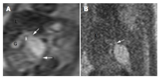
Two weeks later, a fetal MRI (Philips 1.5 T Intera; Erlangen, Germany) was performed to further evaluate the cyst using HASTE sequence (TR 10 ms; TE 100 ms; time of acquisition 12 s; flip angle 90o; field of view 280 mm×250 mm; matrix 256×256). MRI showed a 1.3 cm× 1.2 cm×2.7 cm, ellipsoid, homogeneous cystic lesion extending from the liver hilum posteriorly and inferomedially to the mid-abdomen (Figure 1). This lesion showed hypointensity on T1-weighed and hyperintensity on T2-weighed images. A more detailed anatomical relationship of the cyst to the gall bladder and the biliary tracts was shown. Type 1 choledochal cyst was tentatively diagnosed.

Figure 1
Figure 1 MRI performed at 26 wk’ gestation using HASTE sequence. A: Coronal T2-weighed image showed the choledochal cyst, its tapered ends (arrows), its connection to the cystic duct (arrowhead), and its relationship to the gall bladder (G) and the liver (L); B: Sagittal T2-weighed image showed the posterio-inferior orientation of the choledochal cyst and its connection to the common hepatic duct (arrow).

A female infant weighing 3 890 g was delivered at 39 wk with APGAR scores of 8 and 10 at 1 and 5 min, respectively. Postnatal US at 3 d of age showed further enlargement of the cyst. At 13 d of age, the cyst was excised and a Roux-en-Y hepatico-jejunostomy was performed. Histopathological examination confirmed the diagnosis of a choledochal cyst. The infant was discharged a week later with no jaundice noted thereafter.

DISCUSSION

Choledochal cysts are rare anomalies, characterized by cystic or fusiform dilatation of the intrahepatic and/or extrahepatic biliary tracts. These cysts have been classified into five major types[9]. Cystic dilatation of the common bile duct (type 1 choledochal cyst) comprises 80-90% of the cysts. In affected newborns, about 70% had jaundice but only 25% had a palpable abdominal mass[4]. Potential complications in untreated patients include recurrent cholangitis, pancreatitis, liver cirrhosis, portal hypertension, and malignant transformation[10-12].

Ultrasonography is the primary imaging modality for evaluating the fetus due to its real-time display and non-invasiveness. In cases of choledochal cyst, tubular structures can be identified entering or leaving a right upper quadrant cyst[1,2,4]. However, the choledochal cyst could be missed, if confused with the gall bladder and the umbilical vein. Other common cystic lesions, including hydronephrosis, duodenal duplication, intestinal atresia, mesenteric cyst, and ovarian cyst, may appear similar to a choledochal cyst.

Recent development of ultrafast MR imaging techniques has allowed MRI to become an important complement to US in fetal imaging[5-8]. MRI offers superior soft tissue contrast, larger field of view, and is relatively operator independent. In our case, while the prenatal US showed a subhepatic cyst, the MRI showed additional findings of tapered ends of the cyst, its proximity to the liver hilum, its inferomedial orientation, and characteristic fluid-intensity of the cyst. Also, the relationship between the choledochal cyst, the cystic duct and the dilated gall bladder was clearly depicted. Concerning the differential diagnoses of a right upper quadrant cyst, an umbilical vein may be excluded by the lack of vascular flow void on MRI. Other common cystic lesions, including hydronephrosis, intestinal atresia, mesenteric cyst, and ovarian cyst have locations different from that of a choledochal cyst. Duodenal duplication may not be easily excluded, but it would be less likely to have tapered ends as a choledochal cyst[13]. To our best knowledge, we found two reports of prenatal MRI investigation of choledochal cysts[14,15]. However, the finding of tapered ends of the choledochal cyst, which may be specific to a biliary tract anomaly, was not emphasized in their reports.

In conclusion, owing to its excellent anatomical and contrast resolutions, MRI can clearly delineate a choledochal cyst and may be helpful in differential diagnosis of right upper quadrant cystic lesions. The additional information provided by prenatal MRI, which is complementary to US, may be valuable for family counseling, and may help clinicians manage pregnancy and plan post-natal treatment earlier.

Footnotes

Science Editor Guo SY Language Editor Elsevier HK

References
1.  Elrad H, Mayden KL, Ahart S, Giglia R, Gleicher N. Prenatal ultrasound diagnosis of choledochal cyst. J Ultrasound Med. 1985;4:553-555.  [PubMed]  [DOI]  [Cited in This Article: ]
2.  Frank JL, Hill MC, Chirathivat S, Sfakianakis GN, Marchildon M. Antenatal observation of a choledochal cyst by sonography. AJR Am J Roentgenol. 1981;137:166-168.  [PubMed]  [DOI]  [Cited in This Article: ]  [Cited by in Crossref: 29]  [Cited by in F6Publishing: 30]  [Article Influence: 0.7]  [Reference Citation Analysis (0)]
3.  Howell CG, Templeton JM, Weiner S, Glassman M, Betts JM, Witzleben CL. Antenatal diagnosis and early surgery for choledochal cyst. J Pediatr Surg. 1983;18:387-393.  [PubMed]  [DOI]  [Cited in This Article: ]  [Cited by in Crossref: 73]  [Cited by in F6Publishing: 67]  [Article Influence: 1.6]  [Reference Citation Analysis (0)]
4.  Bancroft JD, Bucuvalas JC, Ryckman FC, Dudgeon DL, Saunders RC, Schwarz KB. Antenatal diagnosis of choledochal cyst. J Pediatr Gastroenterol Nutr. 1994;18:142-145.  [PubMed]  [DOI]  [Cited in This Article: ]  [Cited by in Crossref: 37]  [Cited by in F6Publishing: 36]  [Article Influence: 1.2]  [Reference Citation Analysis (0)]
5.  Simon EM, Goldstein RB, Coakley FV, Filly RA, Broderick KC, Musci TJ, Barkovich AJ. Fast MR imaging of fetal CNS anomalies in utero. AJNR Am J Neuroradiol. 2000;21:1688-1698.  [PubMed]  [DOI]  [Cited in This Article: ]
6.  Johnson IR, Stehling MK, Blamire AM, Coxon RJ, Howseman AM, Chapman B, Ordidge RJ, Mansfield P, Symonds EM, Worthington BS. Study of internal structure of the human fetus in utero by echo-planar magnetic resonance imaging. Am J Obstet Gynecol. 1990;163:601-607.  [PubMed]  [DOI]  [Cited in This Article: ]  [Cited by in Crossref: 38]  [Cited by in F6Publishing: 39]  [Article Influence: 1.1]  [Reference Citation Analysis (0)]
7.  Yamashita Y, Namimoto T, Abe Y, Takahashi M, Iwamasa J, Miyazaki K, Okamura H. MR imaging of the fetus by a HASTE sequence. AJR Am J Roentgenol. 1997;168:513-519.  [PubMed]  [DOI]  [Cited in This Article: ]  [Cited by in Crossref: 162]  [Cited by in F6Publishing: 165]  [Article Influence: 5.9]  [Reference Citation Analysis (0)]
8.  Quinn TM, Hubbard AM, Adzick NS. Prenatal magnetic resonance imaging enhances fetal diagnosis. J Pediatr Surg. 1998;33:553-558.  [PubMed]  [DOI]  [Cited in This Article: ]  [Cited by in Crossref: 178]  [Cited by in F6Publishing: 179]  [Article Influence: 6.6]  [Reference Citation Analysis (0)]
9.  Savader SJ, Benenati JF, Venbrux AC, Mitchell SE, Widlus DM, Cameron JL, Osterman FA. Choledochal cysts: classification and cholangiographic appearance. AJR Am J Roentgenol. 1991;156:327-331.  [PubMed]  [DOI]  [Cited in This Article: ]  [Cited by in Crossref: 54]  [Cited by in F6Publishing: 53]  [Article Influence: 1.6]  [Reference Citation Analysis (0)]
10.  Yamaguchi M. Congenital choledochal cyst. Analysis of 1,433 patients in the Japanese literature. Am J Surg. 1980;140:653-657.  [PubMed]  [DOI]  [Cited in This Article: ]  [Cited by in Crossref: 321]  [Cited by in F6Publishing: 262]  [Article Influence: 5.8]  [Reference Citation Analysis (0)]
11.  Todani T, Tabuchi K, Watanabe Y, Kobayashi T. Carcinoma arising in the wall of congenital bile duct cysts. Cancer. 1979;44:1134-1141.  [PubMed]  [DOI]  [Cited in This Article: ]  [Cited by in F6Publishing: 2]  [Reference Citation Analysis (0)]
12.  Yoshida H, Itai Y, Minami M, Kokubo T, Ohtomo K, Kuroda A. Biliary malignancies occurring in choledochal cysts. Radiology. 1989;173:389-392.  [PubMed]  [DOI]  [Cited in This Article: ]  [Cited by in Crossref: 42]  [Cited by in F6Publishing: 46]  [Article Influence: 1.3]  [Reference Citation Analysis (0)]
13.  Wong AM, Wong HF, Cheung YC, Wan YL, Ng KK, Kong MS. Duodenal duplication cyst: MRI features and the role of MR cholangiopancreatography in diagnosis. Pediatr Radiol. 2002;32:124-125.  [PubMed]  [DOI]  [Cited in This Article: ]  [Cited by in Crossref: 12]  [Cited by in F6Publishing: 13]  [Article Influence: 0.6]  [Reference Citation Analysis (0)]
14.  Mackenzie TC, Howell LJ, Flake AW, Adzick NS. The management of prenatally diagnosed choledochal cysts. J Pediatr Surg. 2001;36:1241-1243.  [PubMed]  [DOI]  [Cited in This Article: ]  [Cited by in Crossref: 57]  [Cited by in F6Publishing: 50]  [Article Influence: 2.1]  [Reference Citation Analysis (0)]
15.  Chen CP, Cheng SJ, Chang TY, Yeh LF, Lin YH, Wang W. Prenatal diagnosis of choledochal cyst using ultrasound and magnetic resonance imaging. Ultrasound Obstet Gynecol. 2004;23:93-94.  [PubMed]  [DOI]  [Cited in This Article: ]  [Cited by in Crossref: 18]  [Cited by in F6Publishing: 10]  [Article Influence: 0.5]  [Reference Citation Analysis (0)]