Copyright
©2005 Baishideng Publishing Group Co.
World J Gastroenterol. Jan 14, 2005; 11(2): 176-181
Published online Jan 14, 2005. doi: 10.3748/wjg.v11.i2.176
Published online Jan 14, 2005. doi: 10.3748/wjg.v11.i2.176
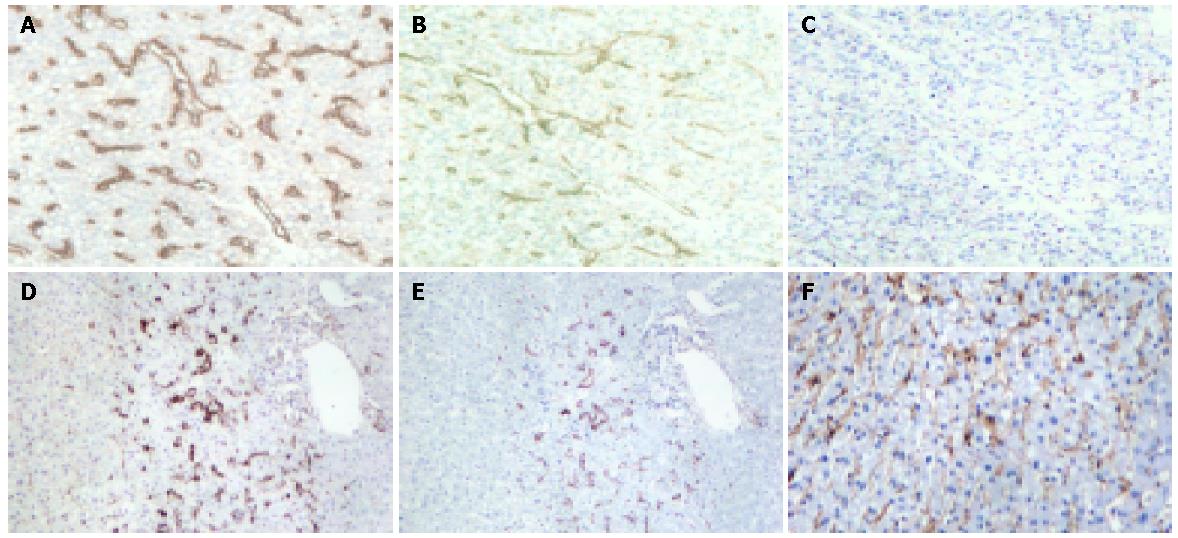
Figure 1 Expression of CD105 and CD34 as shown by the brown staining of the vasculature in tumor and adjacent non-tumorous liver (×100).
A-C are serial sections of tumor tissue, where A shows the CD34 expression, B shows the CD105 expression, and C shows the negative control. D and E are serial sections of adjacent non-tumorous liver, where D shows the CD34 expression, and E shows the CD105 expression. Both D and E illustrate typical focal staining of vessels around the portal vein. F shows a case of diffuse CD105 staining in the non-tumorous liver, whereas such pattern of staining was not seen in the non-tumorous liver by CD34 staining.
- Citation: Ho JW, Poon RT, Sun CK, Xue WC, Fan ST. Clinicopathological and prognostic implications of endoglin (CD105) expression in hepatocellular carcinoma and its adjacent non-tumorous liver. World J Gastroenterol 2005; 11(2): 176-181
- URL: https://www.wjgnet.com/1007-9327/full/v11/i2/176.htm
- DOI: https://dx.doi.org/10.3748/wjg.v11.i2.176


