Copyright
©The Author(s) 2004.
World J Gastroenterol. Dec 1, 2004; 10(23): 3399-3404
Published online Dec 1, 2004. doi: 10.3748/wjg.v10.i23.3399
Published online Dec 1, 2004. doi: 10.3748/wjg.v10.i23.3399
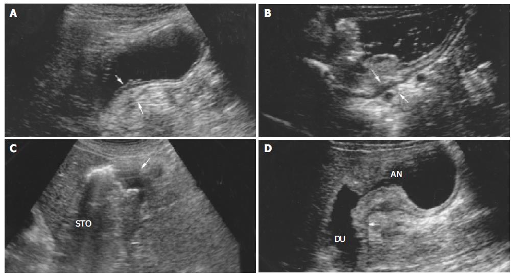
Figure 1 Sonograms of T1-T4 carcinoma.
A: Submucosal carcinoma (T1) in gastric antrum. Arrow indicates segmental thickening of layers 1-3 of the posterior wall, triangle indicates normal layers 4-5. B: T2 carcinoma. The posterior wall of Gastric body is thickening. Arrow indicates the third hyperechoic layer is obliterated and the fourth hypoechoic layer is thickening, triangle indicates normal layer 5. C: T3 carcinoma. Tumor located in the greater curvature of stomach (STO) is hypoechoic with disap-pearance of wall all layer (arrow). D: T4 carcinoma. Sonogram shows tumor located in antrum (AN) infiltrating duodenum. Arrow indicates the segmental wall thickening of duodenal bulb.
- Citation: Liao SR, Dai Y, Huo L, Yan K, Zhang L, Zhang H, Gao W, Chen MH. Transabdominal ultrasonography in preoperative staging of gastric cancer. World J Gastroenterol 2004; 10(23): 3399-3404
- URL: https://www.wjgnet.com/1007-9327/full/v10/i23/3399.htm
- DOI: https://dx.doi.org/10.3748/wjg.v10.i23.3399


