Copyright
©The Author(s) 2003.
World J Gastroenterol. Sep 15, 2003; 9(9): 2114-2116
Published online Sep 15, 2003. doi: 10.3748/wjg.v9.i9.2114
Published online Sep 15, 2003. doi: 10.3748/wjg.v9.i9.2114
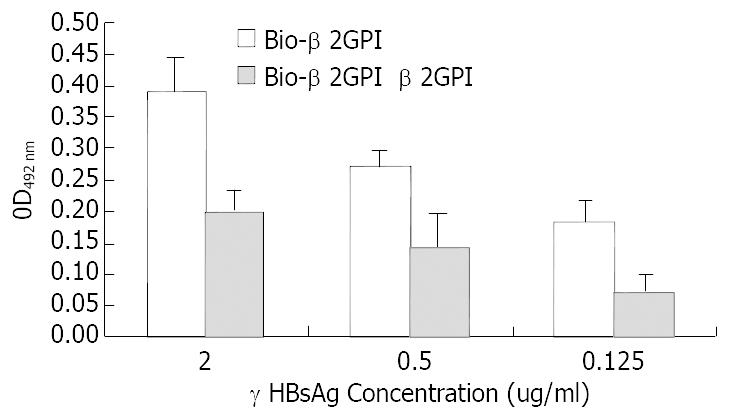
Figure 1 Comparative binding reaction of β2GPI to rHBsAg by ELISA-based determination of protein interaction.
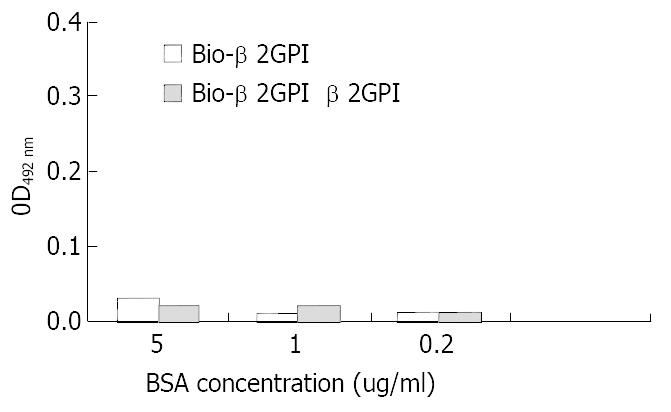
Figure 2 Comparative binding reaction of β2GPI to BSA by ELISA-based determination of protein interaction.
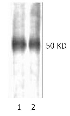
Figure 3 Reduced (lane1) and non-reduced (lane2) human β2GPI (1 μg/lane) was separated with 12% SDS-PAGE and applied to Hybond nitrocellous membrane, blocked with 5% skimmed milk, then probed with biotinylated rHBsAg, color was developed with HRP-avidin in DAB solution.
Single band at -50 kDa was observed, no significant diference in color den-sity was observd, which was further confirmed by digital scaning.
- Citation: Gao PJ, Piao YF, Liu XD, Qu LK, Shi Y, Wang XC, Yang HY. Studies on specific interaction of beta-2-glycoprotein I with HBsAg. World J Gastroenterol 2003; 9(9): 2114-2116
- URL: https://www.wjgnet.com/1007-9327/full/v9/i9/2114.htm
- DOI: https://dx.doi.org/10.3748/wjg.v9.i9.2114


