Copyright
©The Author(s) 2003.
World J Gastroenterol. Sep 15, 2003; 9(9): 2100-2104
Published online Sep 15, 2003. doi: 10.3748/wjg.v9.i9.2100
Published online Sep 15, 2003. doi: 10.3748/wjg.v9.i9.2100
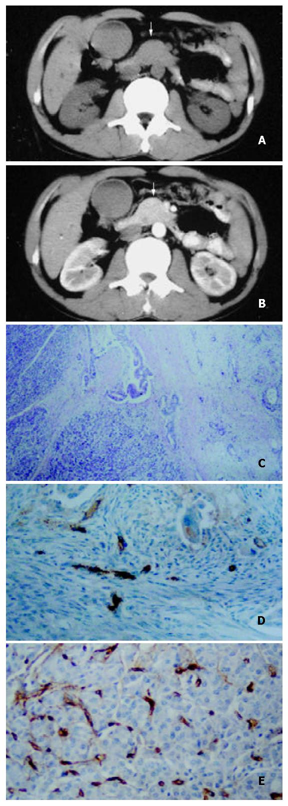
Figure 1 A 52-year-old man with well-differentiated pancreatic head carcinoma.
A: EBCT imaging revealed that bulging pancreatic head (white arrow), density of mass in head and neck were the same as that of pancreatic body; B: EBCT enhancement imaging was 35 s after administration of contrast agent revealed that enhancement of mass in pancreatic head (white arrow) was the same as that of normal pancreatic tissue; C: Hematoxylin-eosin-stained specimen (100 ×) demonstrated the irregularity around tumor cell adeno-tubula structure and residual pancreatic tissue; D: Showing immunohistochemical staining of anti-CD34 antibody (200 ×). CD34-positive cells were counted as microvessels (tan color), MVDs in hot spot area of the neoplastic cells in well-differentiated pancreatic carcinomal; E: High MVDs of residual pancreatic tissue in well differenti-ated pancreatic carcinoma (200 ×).
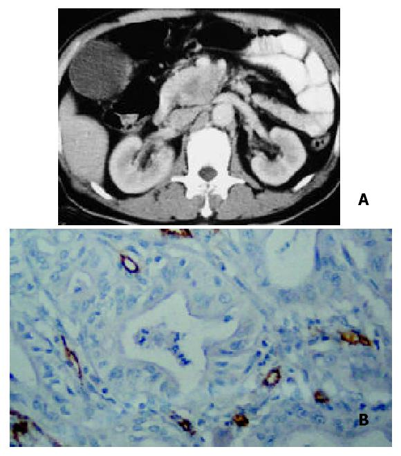
Figure 2 A 52-year-old man with moderately-differentiated pancreatic head carcinoma.
A: Pancreatic-phase helical CT enhancement imaging 35 s after administration of contrast agent revealed that CT enhancement of mass in pancreatic head was slightly lower than that of normal pancreatic tissues. Scarce blood vessel area appeared in the central region of the mass; B: Showing immunohistochemical staining of anti-CD34 antibody (200 ×). CD34-positive cells were counted as microvessels (tan color), moderate MVDs in hot spot area of the neoplastic cells in moderately-differentiated pancreatic carcinoma.
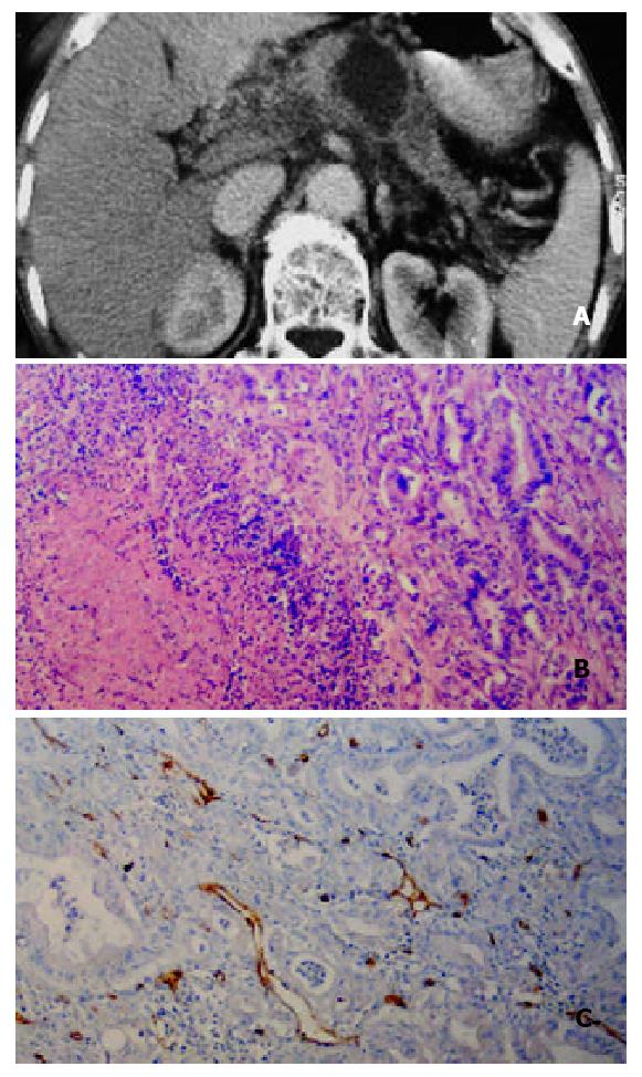
Figure 3 A 59-year-old man with poorly differentiated pancreatic body carcinoma.
A: Helical CT enhancement imag-ing 35 s after administration of contrast agent revealed that pancreatic-phase CT enhancement of mass in pancreatic body was lower than that of surrunding normal pancreatic tissues. Necrotic tissue in the center of the mass was not enhanced. Tumor tissue around the necrotic tissue was slightly enhanced; B:Hematoxylin-eosin-stained specimen (100 ×) demonstrated massive necrosis and decreased re-sidual pancreatic tissue; C: Showing immunohistochemical staining of anti-CD34 antibody (200 ×). CD34-positive cells were seen as microvessels (brown yellow color), more MVDs in hot spot area of the neoplastic cells in poorly-differenti-ated pancreatic carcinoma.
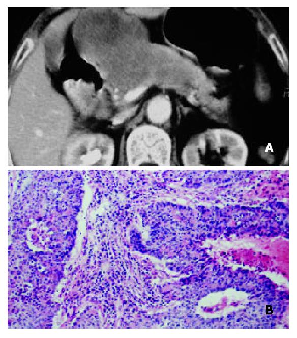
Figure 4 A 70-year-old woman with solid-cystic-papillary well-differentiated pancreatic carcinoma with involvement of ligamentum hepatoduodenale.
A: Helical CT enhancement imaging 35 s after administration of contrast agent revealed that CT enhancement of mass was heterogenously slight low density with low density necrotic region; B: Hematoxylin-eosin-stained specimen (100 ×) demonstrated massive necrosis tissuse and irregular tubula structure in well-differentiated pancreatic carcinoma.
- Citation: Wang ZQ, Li JS, Lu GM, Zhang XH, Chen ZQ, Meng K. Correlation of CT enhancement, tumor angiogenesis and pathologic grading of pancreatic carcinoma. World J Gastroenterol 2003; 9(9): 2100-2104
- URL: https://www.wjgnet.com/1007-9327/full/v9/i9/2100.htm
- DOI: https://dx.doi.org/10.3748/wjg.v9.i9.2100


