Copyright
©The Author(s) 2003.
World J Gastroenterol. Sep 15, 2003; 9(9): 2068-2072
Published online Sep 15, 2003. doi: 10.3748/wjg.v9.i9.2068
Published online Sep 15, 2003. doi: 10.3748/wjg.v9.i9.2068
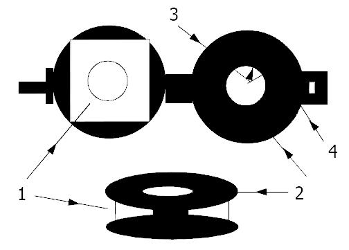
Figure 1 Exhibition map of the tissue-sample holder.
1. Tissue-sample, 2. Tissue-sample holder, 3. Ra = 6 mm 4. Rb = 12 mm.
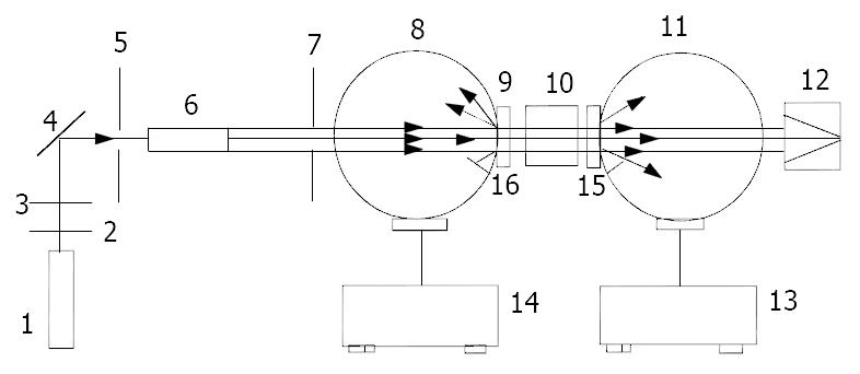
Figure 2 A double-integrating-sphere system was used to determine the optical properties of biological tissues.
1. Laser, 2. Attenuator, 3. Attenuator, 4. Mirror, 5. 2 mm pinhole, 6. Beam expander, 7. 6 mm pinhole, 8. Intergrating sphere I, 9. Sample, 10. Optical trap, 11. Intergrating sphere II, 12. Optical trap, 13. Detector system, 14. Detector system, 15. Baffle, 16. Baffle.
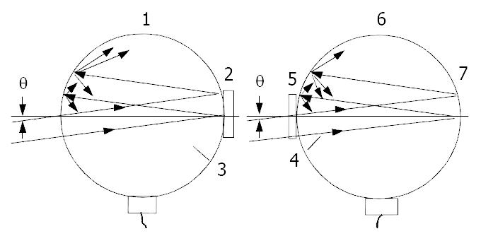
Figure 3 Measuring set of specular reflectance and collimated transmittance of biological tissues.
1. Intergrating sphere I 2. Sample 3. Baffle 4. Baffle 5. Sample 6. Intergrating sphere II 7. Standard reference plate θ = 3o.
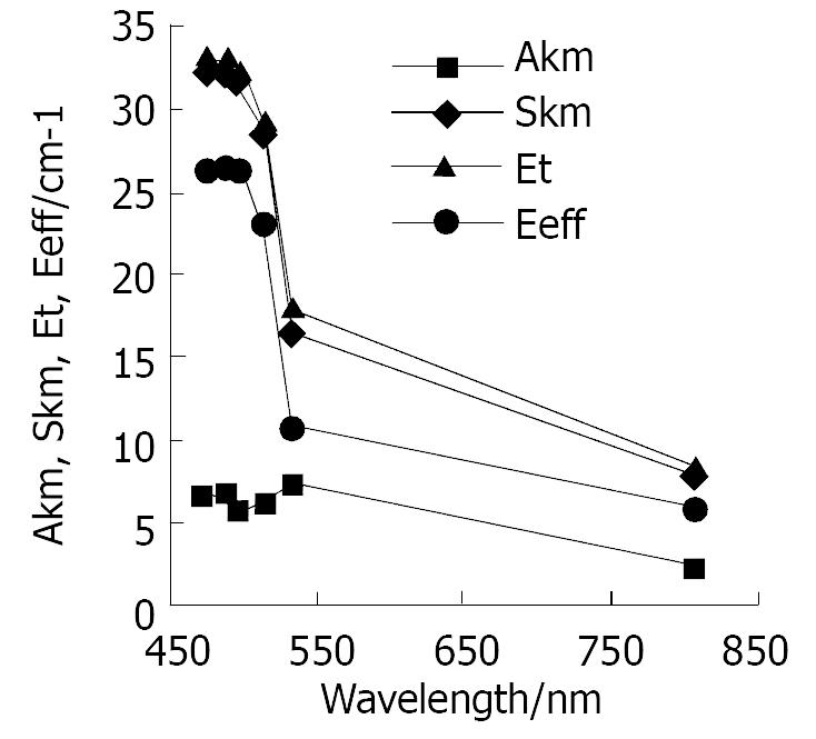
Figure 4 Broken line graphs of AKM-λ, SKM-λ, Et-λ, Seff-λ of human normal small intestine tissue.
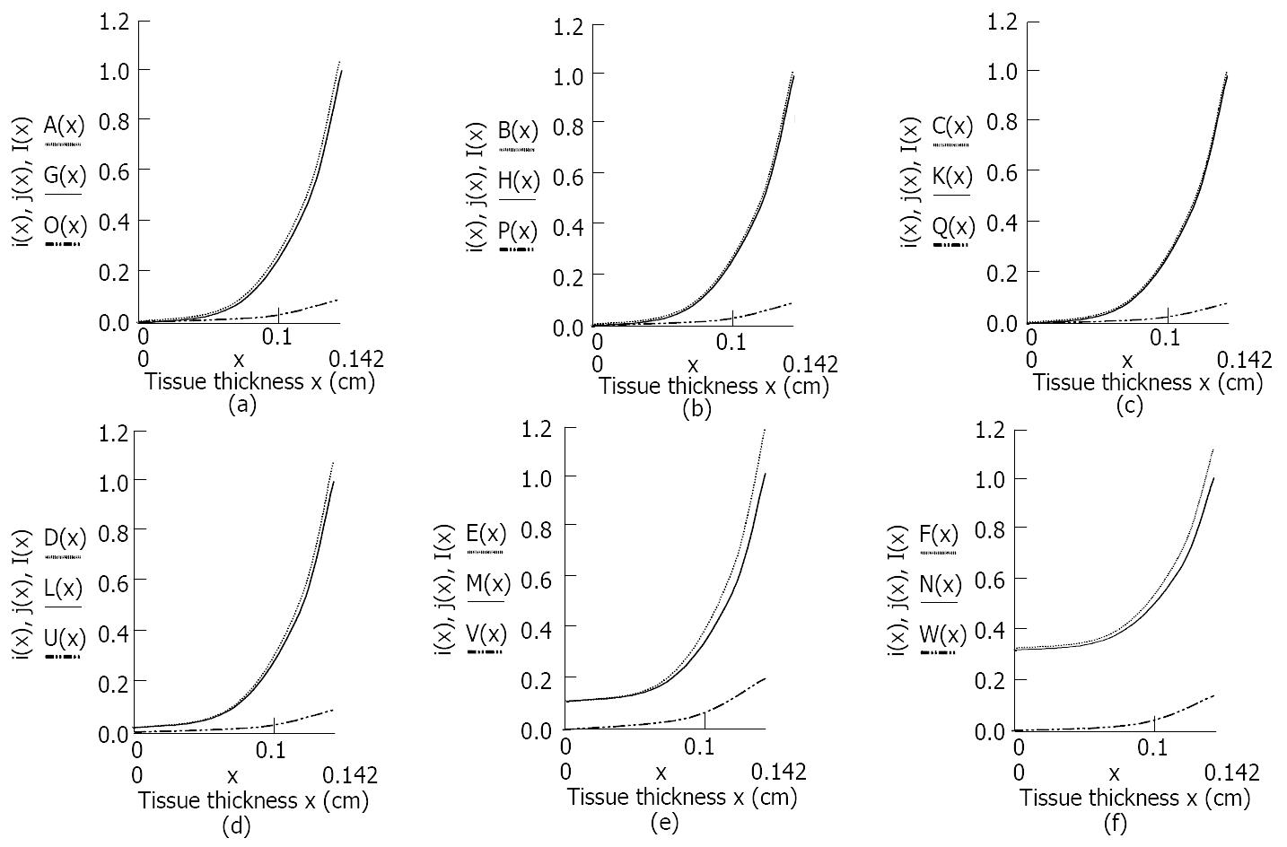
Figure 5 Light distribution of i (x), j (x), I (x) of human normal small intestine tissue in Kubelka-Munk two-flux model changed with tissue thickness at six different wavelengths of laser radiation.
(a) A(x), G(x) and O(x) respectively represented the forward and backward, and the total scattered photon fluxes of the tissue at 476.5 nm laser irradiation. (b) B(x), H(x) and P(x) respectively represented the forward, backward and the total scattered photon fluxes of the tissue at 488 nm laser irradiation. (c) C(x), K(x) and Q(x) respectively represented the forward, backward and the total scattered photon fluxes of the tissue at 496.5 nm laser irradiation. (d) D(x), L(x) and U(x) respectively represented the forward, backward and the total scattered photon fluxes of the tissue at 514.5 nm laser irradiation. (e) E(x), M(x) and V(x) respectively represented the forward, backward and the total scattered photon fluxes of the tissue at 532 nm laser irradiation. (f) F(x), N(x) and W(x) respectively represented the forward, backward and the total scattered photon fluxes of the tissue at 808 nm laser irradiation.
-
Citation: Wei HJ, Xing D, Wu GY, Jin Y, Gu HM. Optical properties of human normal small intestine tissue determined by Kubelka-Munk method
in vitro . World J Gastroenterol 2003; 9(9): 2068-2072 - URL: https://www.wjgnet.com/1007-9327/full/v9/i9/2068.htm
- DOI: https://dx.doi.org/10.3748/wjg.v9.i9.2068


