Copyright
©The Author(s) 2003.
World J Gastroenterol. Aug 15, 2003; 9(8): 1719-1724
Published online Aug 15, 2003. doi: 10.3748/wjg.v9.i8.1719
Published online Aug 15, 2003. doi: 10.3748/wjg.v9.i8.1719
Figure 1 Nucleotide and Predicted Amino Acid Sequence of the APMCF1 cDNA and sequence alignment of APMCF1 wth the protein from other species.
A: The full-length gene sequence and amino acids sequence of APMCF1; B: Alignment was done by using the Align software in Vector NTI. Dark gray background shows identity with at lease five aligned residues in different species. The gray region represents the sequence conserved among different species.
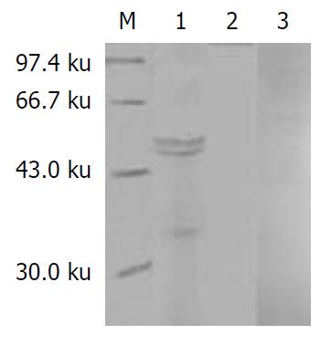
Figure 2 Immunoblotting of APMCF1 in GST-APMCF1 (56 ku) using antiserum, normal rabbit serum before immunization and antiserum preabsorbed with GST-APMCF1.
M: Protein marker; 1: antiserum; 2: rabbit serum before immune from GST-APMCF1; 3: antiserum absorbed in the peptides used for im-munization (control antibody).
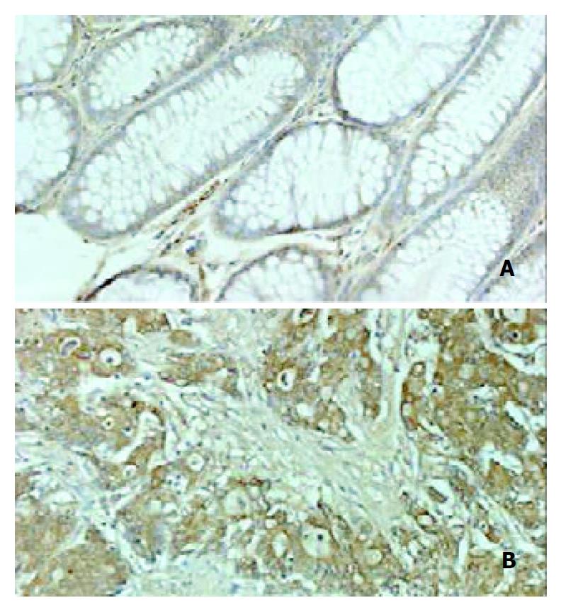
Figure 3 Expression of AMPCF1 in normal colon tissue and colon cancer ( × 200).
A. Normal colon tissue; B. Colon cancer.
- Citation: Yan W, Wang WL, Zhu F, Chen SQ, Li QL, Wang L. Isolation of a novel member of small G protein superfamily and its expression in colon cancer. World J Gastroenterol 2003; 9(8): 1719-1724
- URL: https://www.wjgnet.com/1007-9327/full/v9/i8/1719.htm
- DOI: https://dx.doi.org/10.3748/wjg.v9.i8.1719


