Copyright
©The Author(s) 2003.
World J Gastroenterol. Aug 15, 2003; 9(8): 1667-1674
Published online Aug 15, 2003. doi: 10.3748/wjg.v9.i8.1667
Published online Aug 15, 2003. doi: 10.3748/wjg.v9.i8.1667
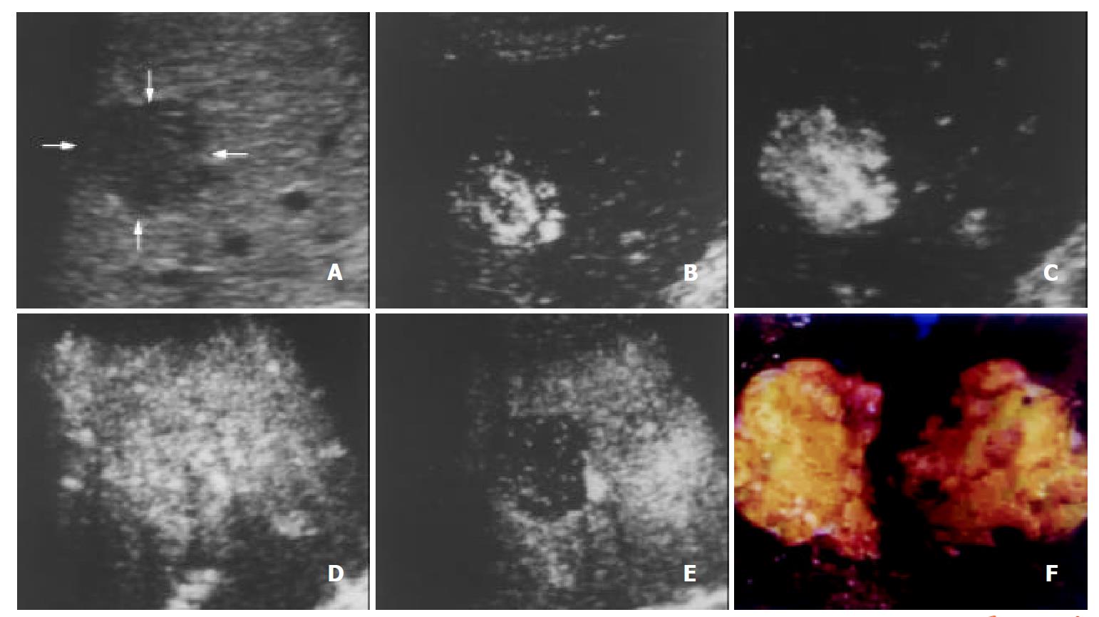
Figure 1 A 47-year-old man with hepatocellular carcinoma.
A. Intercostal precontrast conventional sonography exhibits a hypoechogenic lesion (arrows) of 2.7 cm in diameter. B-E. Contrast-enhanced C-cube gray scale sonography at 23 s (B), 28 s (C) and 43 s (D) after injection of Levovist shows that intranodular signals enhance gradually and earlier than the liver parenchyma, and enhancement decreases rapidly in the portal venous phase (110 s, E). This suggests hypervascularity of hepatocellular carcinoma with an early enhancement and early wash-out of contrast. F. Gross specimen of the resected tumor exhibits a gray fish-like profile and suggests the typical appearance of hepatocellular carcinoma.
Figure 2 A 48-year-old man with metastasis of nasopharyngeal carcinoma.
A. Intercostal section of precontrast conventional sonography exhibits a hypoechogenic lesion (arrows) with a diameter of 3.5 cm. B. Contrast-enhanced C-cube gray scale sonography at 24 s after injection of Levovist shows that peripheral enhancement appears at the same time with the liver parenchyma. C. Intranodular enhancement decreases at 107 s in the portal venous phase, earlier than that in the liver parenchyma.
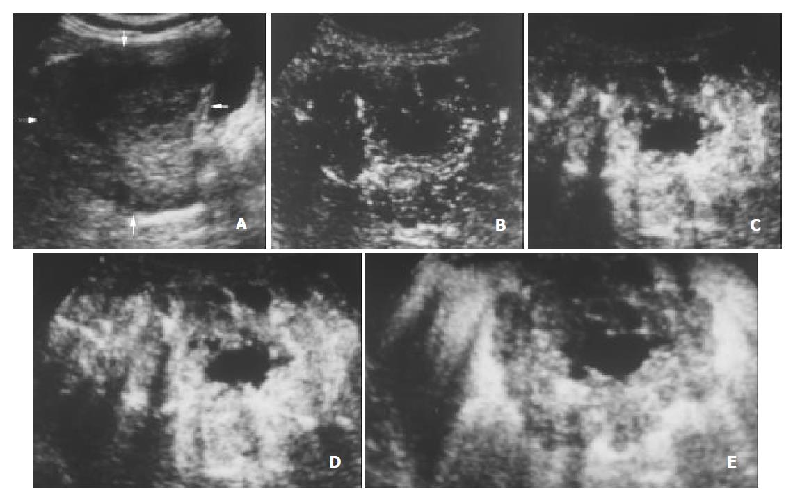
Figure 3 A 62-year-old woman with cholangiocarcinoma.
A. Subcostal section of precontrast conventional sonography shows a hypoechogenic lesion (arrows) in segment V of the liver with a diameter of 8.0 cm. B-E. Contrast-enhanced C-cube gray scale sonography at 16 s (B), 20 s (C) and 25 s (D) after injection of Levovist shows that intranodular signals enhance gradually and earlier than the liver parenchyma, and enhancement decreases rapidly in the portal venous phase (78 s, E). Histopathology of US-guided percutaneous biopsy reveals cholangiocarcinoma of the liver.
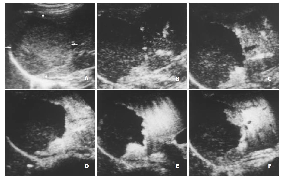
Figure 4 A 45-year-old female with hemangioma.
A. Intercostal section of precontrast conventional sonography exhibits a hypoechogenic lesion (arrows) of 6.5 cm in diameter. B. Contrast-enhanced C-cube gray scale sonography at 23 s (B) after injection of Levovist shows that no microbubble signal appears in the lesion while enhancement in the liver parenchyma begins. C-F. C-cube gray scale sonography demonstrates gradual peripheral enhancement at 32 s (C), 47 s (D), 113 s (E) and 370 s (F) with a long enhancement duration.
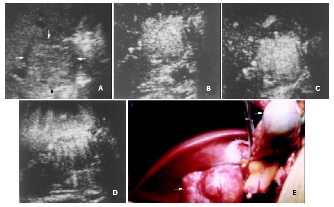
Figure 5 A 29-year-old man with focal nodular hyperplasia.
A. Intercostal section of precontrast conventional sonography exhibits an isoechoic lesion (arrows) in segment V of the liver with a diameter of 4.8 cm. B-D. Contrast-enhanced C-cube gray scale sonography at 49 s (B) and 60 s (C) after injection of Levovist shows that intranodular signals enhance earlier than the liver parenchyma, and enhancement sustains in the portal venous phase until the delayed phase (140 s, D), suggestive of slow wash-out of contrast in the lesion. E. Appearance of the lesion and its surrounding organs in laparotomy. The lesion (arrow) protrudes from the liver with a smooth capsule while the gallbladder (arrowhead) is lifted with hemostatic forceps. Histopathology of the tumor revealed focal nodular hyperplasia.
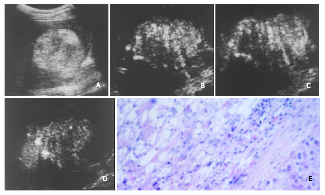
Figure 6 A 51-year-old female with angiomyolipoma.
A. Subcostal section of precontrast conventional sonography exhibits a hyperechogenic lesion of 7.3 cm in diameter. B-D. Contrast-enhanced C-cube gray scale sonography at 54 s (B) and 110 s (C) after injection of Levovist shows that intranodular signals enhance earlier than the liver parenchyma with an inhomogenous enhance-ment pattern, and enhancement decreases in the delayed phase (206 s, D). E. Photomicrography shows defuse sheets of adipocytes and muscle cells adjacent to the noncirrhotic liver tissue, presenting a typical appearance of angiomyolipoma (HE 400).
- Citation: Wang WP, Ding H, Qi Q, Mao F, Xu ZZ, Kudo M. Characterization of focal hepatic lesions with contrast-enhanced C-cube gray scale ultrasonography. World J Gastroenterol 2003; 9(8): 1667-1674
- URL: https://www.wjgnet.com/1007-9327/full/v9/i8/1667.htm
- DOI: https://dx.doi.org/10.3748/wjg.v9.i8.1667


