Copyright
©The Author(s) 2003.
World J Gastroenterol. Jul 15, 2003; 9(7): 1477-1481
Published online Jul 15, 2003. doi: 10.3748/wjg.v9.i7.1477
Published online Jul 15, 2003. doi: 10.3748/wjg.v9.i7.1477
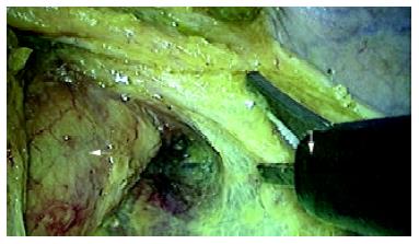
Figure 1 The "lateral" ligaments (→) of rectum containing middle rectal artery or its branches, and mesorectum (←) were dissected completely with a harmonic scalpel (↓).
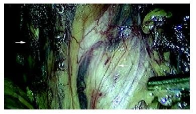
Figure 2 The left pelvic splanchnic nerves were preserved intact as far as possible.
Inset shows the inferior hypogastric nerve nerve fibers (←) and the ureter (→).
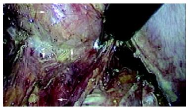
Figure 3 Denonvilliers fascia (↓)was dissected along the space (↑) between the posterior wall of vagina (→) and the rectum (←).
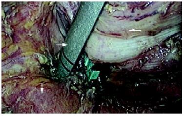
Figure 4 The cross clamping of the rectum (←) was performed 1.
5~3.5cm below the tumor with endo-cutter (→). Pelvic floor 'muscularization' was shown (↓).
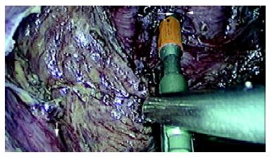
Figure 5 The puncturing cone (→) of the circular stapler pricked through the midpoint of occluding line of the distal rectum(←).
Levator ani muscles were exposed (↓).
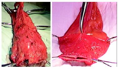
Figure 6 The dorsal mesorectum (→) and distal mesorectum (↓) of the rectal specimen were shown (6a); The anterior side of the specimen and distal margin (←) were shown (6b).
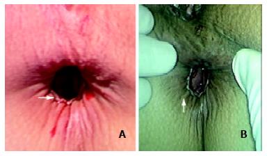
Figure 7 The anastomotic ring (→) could be shown easily in the patient receiving colo-anal anastomosis (a); Satisfactory contractive function of the saved anus (↑) was achieved in the patients receiving laparoscopic TME with anal sphincter preservation at the first day after operation (b).
- Citation: Zhou ZG, Wang Z, Yu YY, Shu Y, Cheng Z, Li L, Lei WZ, Wang TC. Laparoscopic total mesorectal excision of low rectal cancer with preservation of anal sphincter: A report of 82 cases. World J Gastroenterol 2003; 9(7): 1477-1481
- URL: https://www.wjgnet.com/1007-9327/full/v9/i7/1477.htm
- DOI: https://dx.doi.org/10.3748/wjg.v9.i7.1477


