Copyright
©The Author(s) 2003.
World J Gastroenterol. Jul 15, 2003; 9(7): 1455-1459
Published online Jul 15, 2003. doi: 10.3748/wjg.v9.i7.1455
Published online Jul 15, 2003. doi: 10.3748/wjg.v9.i7.1455
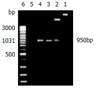
Figure 1 Recombinant plasmid pGEX-4T-1(His)6C-ARL was identified by restrictive endonuclease and polymerase chain reaction (1.
5% agarose electrophoresis). Lane 1: The recombinant plasmid pGEX-4T-1(His)6C-ARL digested by EcoR I only, about 5.9 kb fragment could be seen; Lane 2: The recombinant plasmid pGEX-4T-1(His)6 C-ARL digested by EcoR I and Xho I, two fragments of 5.0 kb and 950 bp could be seen; Lane 3: pGEX-4T-1(His)6C-ARL being template, 950 bp was amplified by polymerase chain reaction; Lane 4: positive control of PCR; Lane 5: negative control of PCR; Lane 6: GeneRulerTM 100 bp DNA ladder plus.
Figure 2 Recombinant plasmid pQE-30-ARL was identified by restrictive endonuclease (1.
5% agarose electrophoresis). Lane 1: 3.4 kb and 0.7 kb and 0.3 kb fragments of pQE-30-ARL digested by BamHI and Hind III together; Lane 2: 0.7 kb and 3.8 kb fragments of pQE-30-ARL digested by BamHI only; Lane 3: 4.5 kb fragment of pQE-30-ARL digested by Hind III alone; Lane 4: GeneRulerTM 100 bp DNA ladder plus.
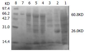
Figure 3 The transformed pGEX-4T-1(His)6C-ARL BL21 induced by 0.
05 mM isopropyl-1-thio-β-D-galactopyranoside. Lane 1: BL21 transformed with pGEX-4T-1(His)6C, no IPTG induced; Lane 2: BL21 transformed with pGEX-4T-1(His)6C, IPTG induced, GST (26KD) was expressed; Lane 3: BL21 transformed with pGEX-4T-1(His)6C-ARL, no IPTG induced; Lane 4: BL21 transformed with pGEX-4T-1(His)6C-ARL, IPTG induced, recombinant protein ARL-GST (about 60.8 KD) was expressed; Lane 5: supernatant; Lane 6: pellet; Lane 7: purified ARL-GST; Lane 8: mid-range protein molecular weight markers.
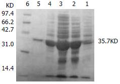
Figure 4 The transformed pQE-30-ARL BL21 induced by 0.
05 mM isopropyl-1-thio-β-D-galactopyranoside. Lane 1: BL21 transformed with pQE-30-ARL, no IPTG induced; Lane 2: BL21 transformed with pQE-30-ARL, IPTG induced, ARL-(His)6(35.7 KD) could express; Lane 3: supernatant; Lane 4: pellet; Lane 5: purified ARL-(His)6; Lane 6: mid-range protein molecular weight markers.
Figure 5 The gel eluted with 0.
1 M glycine (pH2.8).
Figure 6 Proteins of all bacteria described were tested by Western blotting using polyclonal antibodies against ARL-1.
Lane 1: purified ARL-GST; Lane 2: BL21 transformed with pGEX-4T-1(His)6C-ARL; Lane 3: BL21 transformed with pGEX-4T-1(His)6C; Lane 4: purified ARL-(His)6; Lane 5: BL21 transformed with pQE-30-ARL; Lane 6: BL21 transformed with pET-CTF; Lane 7: BL21; Lane 8: Rainbow markers.
- Citation: Jin JF, Yuan LD, Liu L, Zhao ZJ, Xie W. Preparation and characterization of polyclonal antibodies against ARL-1 protein. World J Gastroenterol 2003; 9(7): 1455-1459
- URL: https://www.wjgnet.com/1007-9327/full/v9/i7/1455.htm
- DOI: https://dx.doi.org/10.3748/wjg.v9.i7.1455


