Copyright
©The Author(s) 2003.
World J Gastroenterol. Jul 15, 2003; 9(7): 1421-1426
Published online Jul 15, 2003. doi: 10.3748/wjg.v9.i7.1421
Published online Jul 15, 2003. doi: 10.3748/wjg.v9.i7.1421
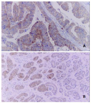
Figure 1 Immunohistochemical staining of COX-2 protein in gastric carcinoma.
Immunoreactivity for COX-2 protein was present in the cytoplasm of tumor cells, smooth muscle cells (A, × 200), and surrounding glands (B, × 100).
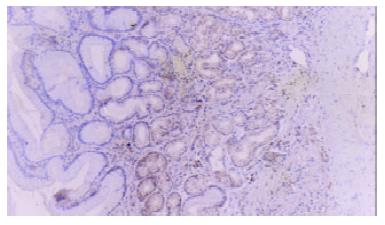
Figure 2 Positive immunostaining for COX-2 was observed in some CAG specimens (× 100).
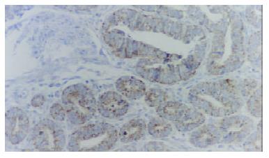
Figure 3 Immunohistochemical staining for VEGF in gastric carcinoma.
VEGF was mainly localized on the membrane of the carcinoma cells or in the cytoplasm (× 200).
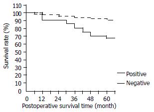
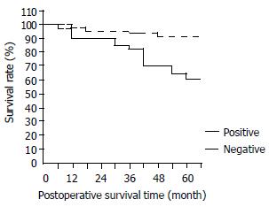
Figure 4 Kaplan-Meier survival curves of patients with gastric carcinoma with regard to COX-2 expression (positive and negative), χ2 = 7.
56, P < 0.01.
Figure 5 Kaplan-Meier survival curves of patients with gastric carcinoma with regard to VEGF expression (positive and negative), χ2 = 16.
51, P < 0.01.
- Citation: Shi H, Xu JM, Hu NZ, Xie HJ. Prognostic significance of expression of cyclooxygenase-2 and vascular endothelial growth factor in human gastric carcinoma. World J Gastroenterol 2003; 9(7): 1421-1426
- URL: https://www.wjgnet.com/1007-9327/full/v9/i7/1421.htm
- DOI: https://dx.doi.org/10.3748/wjg.v9.i7.1421


