Copyright
©The Author(s) 2003.
World J Gastroenterol. May 15, 2003; 9(5): 894-898
Published online May 15, 2003. doi: 10.3748/wjg.v9.i5.894
Published online May 15, 2003. doi: 10.3748/wjg.v9.i5.894
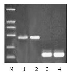
Figure 1 Semiquantitative RT-PCR for ZNRD1 and β2-microglobulin.
M: Marker (DL2000); Lane 1, 3: SGC7901 cells; 2, 4: SGC7901/VCR cells.
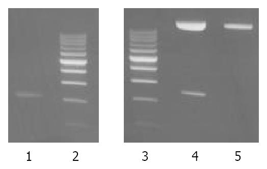
Figure 2 Electrophoresis of PCR product and pUCm-T-ZNRD1 digested with enzymes.
Lane 1: PCR product; Lane 2, 3: Marker (200 bp); Lane 4: recombinant of pUCm-T-ZNRD1 cleaved by EcoR V and Xba I; Lane 5: recombinant of pUCm-T-ZNRD1.
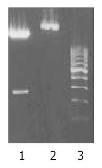
Figure 3 Electrophoresis identification of pcDNA3.
1-anti ZNRD1. Lane 1: pcDNA3.1-anti ZNRD1/EcoR V + BamH I; Lane 2: pcDNA3.1-anti ZNRD1; Lane 3: Marker (200 bp).
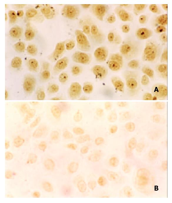
Figure 4 Detection of ZNRD1 expression in cells by immunocytochemical staining.
× 200. A: SGC7901/VCR; B: anti ZNRD1-SGC7901/VCR.
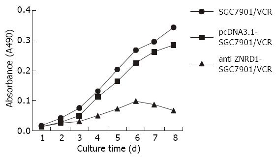
Figure 5 Growth curve of cells.
- Citation: Zhang YM, Zhao YQ, Pan YL, Shi YQ, Jin XH, Yi H, Fan DM. Effect of ZNRD1 gene antisense RNA on drug resistant gastric cancer cells. World J Gastroenterol 2003; 9(5): 894-898
- URL: https://www.wjgnet.com/1007-9327/full/v9/i5/894.htm
- DOI: https://dx.doi.org/10.3748/wjg.v9.i5.894


