Copyright
©The Author(s) 2003.
World J Gastroenterol. Feb 15, 2003; 9(2): 368-372
Published online Feb 15, 2003. doi: 10.3748/wjg.v9.i2.368
Published online Feb 15, 2003. doi: 10.3748/wjg.v9.i2.368
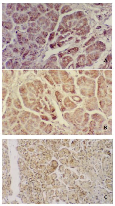
Figure 1 Immunohistochemical staining.
A, B and C: The immunoreactivity of E-cadherin and alpha-, beta-catenin was expressed by normal ductal and acinar cells with strong membranous staining on the intercellular border in the same specimen. × 100.
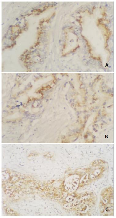
Figure 2 Immunohistochemical staining.
A, B and C: E-cadherin and alpha-, beta-catenin expression mainly were in cytoplasm, whereas membranous staining reduced or disappeared in pancreatic cancer tissue. × 100.
- Citation: Li YJ, Ji XR. Relationship between expression of E-cadherin-catenin complex and clinicopathologic characteristics of pancreatic cancer. World J Gastroenterol 2003; 9(2): 368-372
- URL: https://www.wjgnet.com/1007-9327/full/v9/i2/368.htm
- DOI: https://dx.doi.org/10.3748/wjg.v9.i2.368


