Copyright
©The Author(s) 2003.
World J Gastroenterol. Dec 15, 2003; 9(12): 2873-2875
Published online Dec 15, 2003. doi: 10.3748/wjg.v9.i12.2873
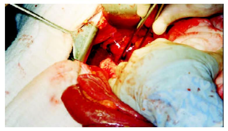
Published online Dec 15, 2003. doi: 10.3748/wjg.v9.i12.2873
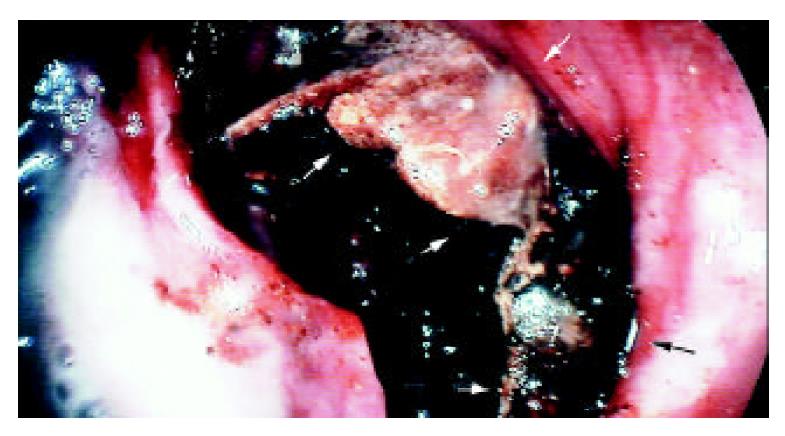
Figure 1 Endoscopic appearance of a large duodenal stone (black arrows) causing complete obstruction.
Note the irregu-lar edges of the stone (white arrows), which indicate its fragmentation.
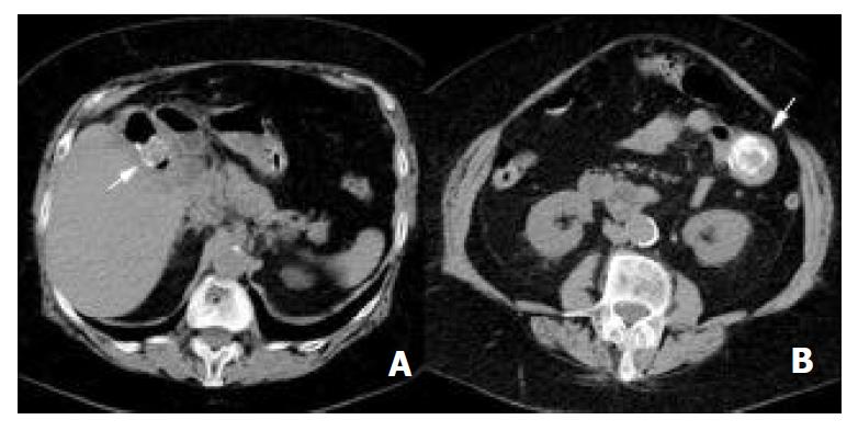
Figure 2 CT shows: A: A large, 5×3×3 cm, intraluminal stone (arrow) in the proximal jejunum, B: Another stone in the duode-nal bulb (arrow).
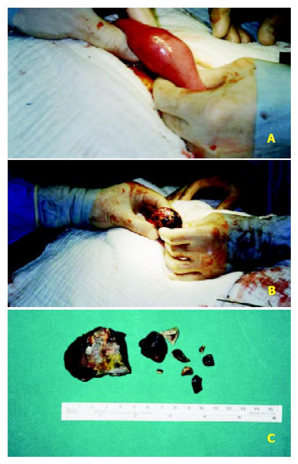
Figure 3 Intraoperative views: A: The obstructed proximal jejunal segment, note a large intraluminal stone causing complete intestinal obstruction at this level, B: Removal of the stone with enterotomy, C: Macroscopic view of the fragmented gallstone which had a very hard outer shell with a soft core.
Figure 4 After the adhesions between the gallbladder (arrow) and the adjacent organs were dissected, cholecystoduodenal fistula (arrow head) was broken down and then the retained stone was removed.
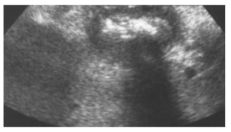
Figure 5 Intraoperative ultrasound revealed that the suspicious stone was in the gallbladder instead of the duodenal lumen.
- Citation: Gencosmanoglu R, Inceoglu R, Baysal C, Akansel S, Tozun N. Bouveret’s syndrome complicated by a distal gallstone Ileus. World J Gastroenterol 2003; 9(12): 2873-2875
- URL: https://www.wjgnet.com/1007-9327/full/v9/i12/2873.htm
- DOI: https://dx.doi.org/10.3748/wjg.v9.i12.2873


