Copyright
©The Author(s) 2003.
World J Gastroenterol. Dec 15, 2003; 9(12): 2870-2872
Published online Dec 15, 2003. doi: 10.3748/wjg.v9.i12.2870
Published online Dec 15, 2003. doi: 10.3748/wjg.v9.i12.2870
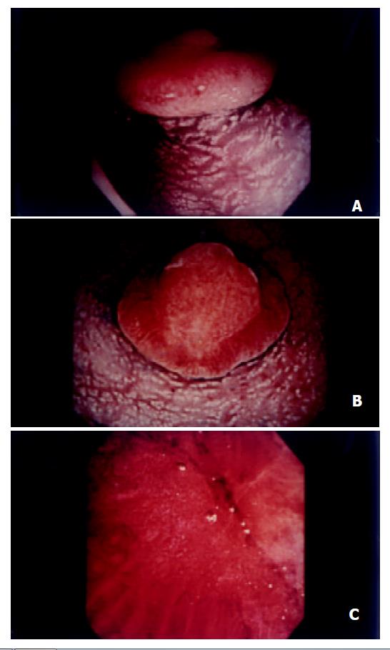
Figure 1 A: Pedunculated polypoid lesion presenting a mush-room appearance in the rectum; B: A round and shallow ero-sion in the center and a marked mucosal bulge at the edge of polyp; C: A non-structural pit pattern in the center with elon-gated pits at the edge revealed in magnifying endoscopy.
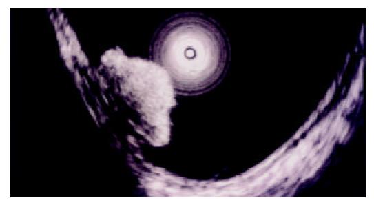
Figure 2 An endoscopic ultrasonography demonstrated ho-mogeneous hypoechoic mass.
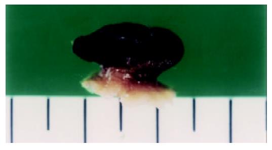
Figure 3 Gross appearance of excised polyp showing a white-yellowish and solid tumor, measuring 13×10 mm with a pedicle.
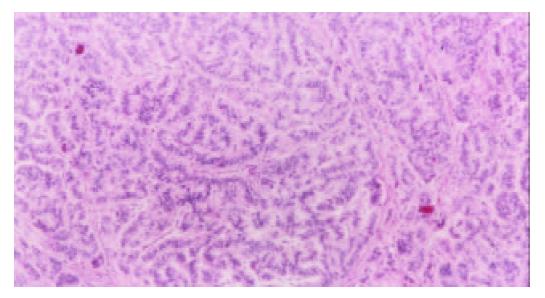
Figure 4 Histology of tumor showing small uniform cells ar-ranged in small nests and cords with an anastomosing ribbon-like pattern in the submucosal layer.
- Citation: Hamada H, Shikuwa S, Wen CY, Isomoto H, Nakao K, Miyashita K, Daikoku M, Yano K, Ito M, Mizuta Y, Chen LD, Xu ZM, Murata I, Kohno S. Pedunculated rectal carcinoid removed by endoscopic mucosal resection: a case report . World J Gastroenterol 2003; 9(12): 2870-2872
- URL: https://www.wjgnet.com/1007-9327/full/v9/i12/2870.htm
- DOI: https://dx.doi.org/10.3748/wjg.v9.i12.2870


