Copyright
©The Author(s) 2003.
World J Gastroenterol. Dec 15, 2003; 9(12): 2813-2816
Published online Dec 15, 2003. doi: 10.3748/wjg.v9.i12.2813
Published online Dec 15, 2003. doi: 10.3748/wjg.v9.i12.2813
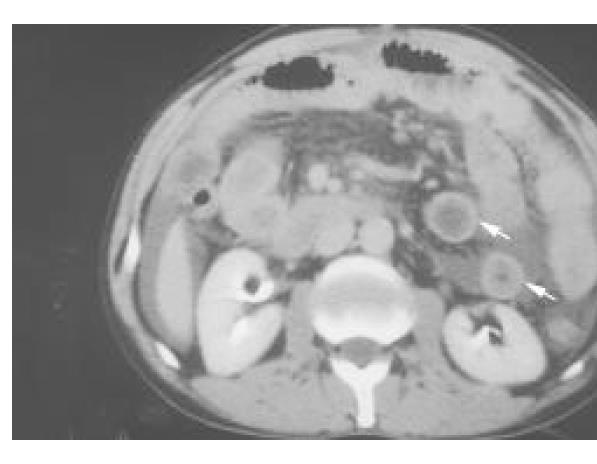
Figure 1 Abdominal computed tomography with intravenous contrast medium showing general thickening in the small bowel wall (white arrows), characteristic of the distribution of eosinophilic gastroenteritis.
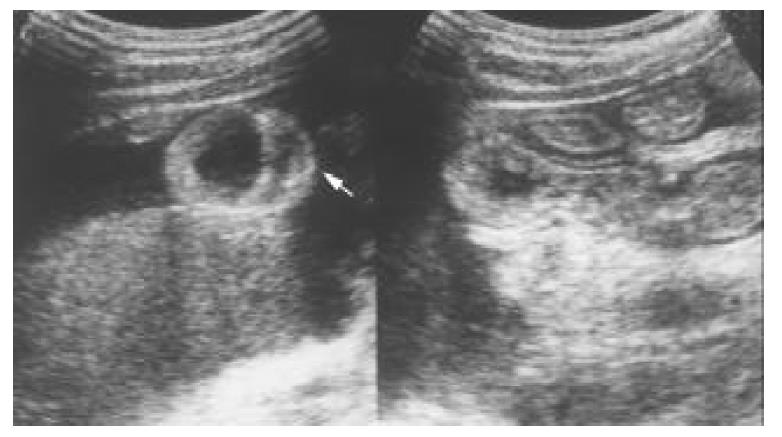
Figure 2 Transverse sonography of the proximal small bowel in the right subcostal area showing general thickening of the wall (white arrow) and ascites.
- Citation: Chen MJ, Chu CH, Lin SC, Shih SC, Wang TE. Eosinophilic gastroenteritis: Clinical experience with 15 patients. World J Gastroenterol 2003; 9(12): 2813-2816
- URL: https://www.wjgnet.com/1007-9327/full/v9/i12/2813.htm
- DOI: https://dx.doi.org/10.3748/wjg.v9.i12.2813


