Copyright
©The Author(s) 2003.
World J Gastroenterol. Dec 15, 2003; 9(12): 2764-2767
Published online Dec 15, 2003. doi: 10.3748/wjg.v9.i12.2764
Published online Dec 15, 2003. doi: 10.3748/wjg.v9.i12.2764
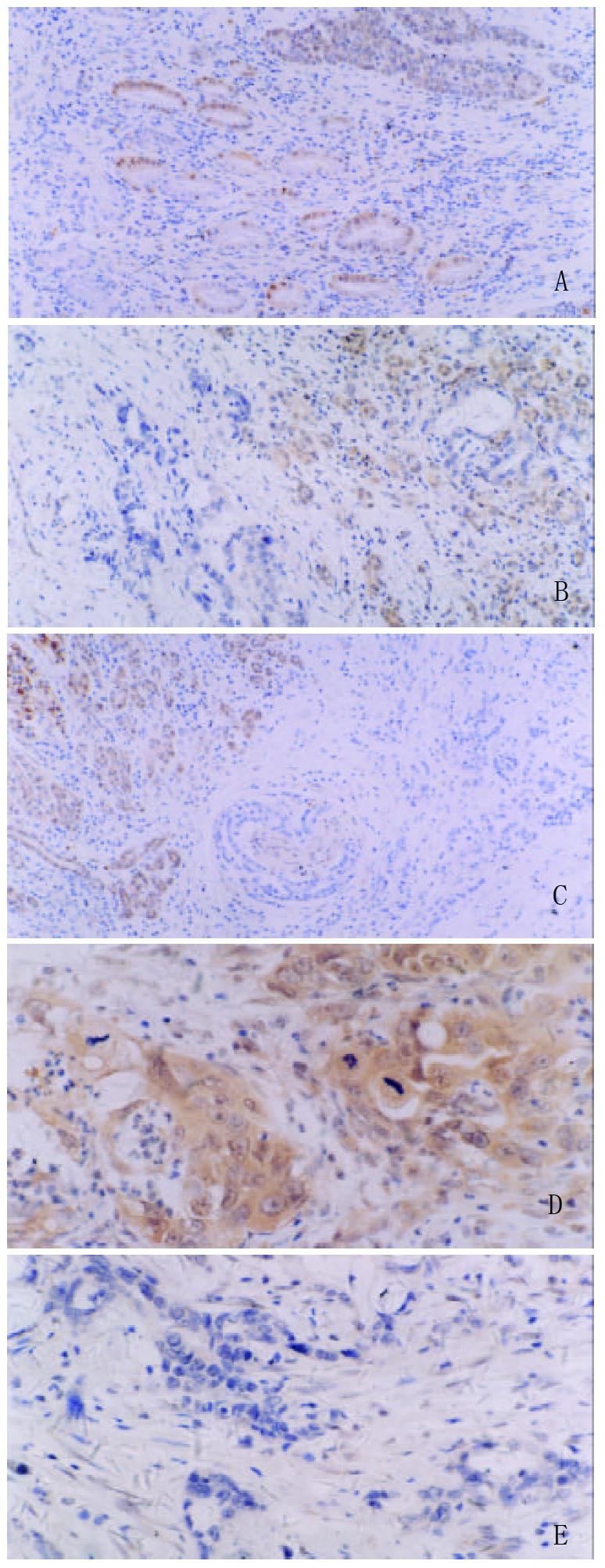
Figure 1 Representative immunostaining results of DPC4 in pancreatic carcinoma (A-E).
Positive cells were stained dark brown in the nuclei and/or cytoplasms (A-D). A: Well-differentiated pancreatic carcinoma showed DPC4 expression. hematoxylin counterstain. original magnification, ×100. B: Well-differentiated pancreatic carcinoma showed loss of DPC4 expression (left), whereas the adjacent normal pancre-atic tissue had DPC4 expression (right). hematoxylin counterstain. original magnification, ×200. C: Moderately-differentiated pancreatic carcinoma showed loss of DPC4 expression (right), whereas the adjacent normal pan-creatic tissue had DPC4 expression (left). Vortex in the middle shows invasion of pancreatic nerve. hematoxylin counterstain. original magnification, ×160. D: Poorly-differentiated pancreatic carcinoma showed DPC4 expression. Hematoxylin counterstain. original magnification, ×400. E: Poorly-differentiated pancreatic carcinoma showed loss of DPC4 expression. hematoxylin counterstain. original magnification, ×400.
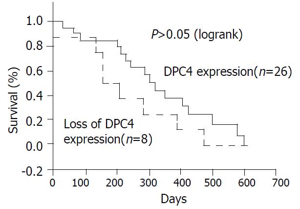
Figure 2 Kaplan-Meier survival curves comparing patients with DPC4 expression and patients with loss of DPC4 expression.
Although patients with DPC4 expression had a higher survival rate than those with loss of DPC4 expression, the difference did not reach ang statistical significance.
- Citation: Hua Z, Zhang YC, Hu XM, Jia ZG. Loss of DPC4 expression and its correlation with clinicopathological parameters in pancreatic carcinoma. World J Gastroenterol 2003; 9(12): 2764-2767
- URL: https://www.wjgnet.com/1007-9327/full/v9/i12/2764.htm
- DOI: https://dx.doi.org/10.3748/wjg.v9.i12.2764


