Copyright
©The Author(s) 2003.
World J Gastroenterol. Nov 15, 2003; 9(11): 2632-2634
Published online Nov 15, 2003. doi: 10.3748/wjg.v9.i11.2632
Published online Nov 15, 2003. doi: 10.3748/wjg.v9.i11.2632
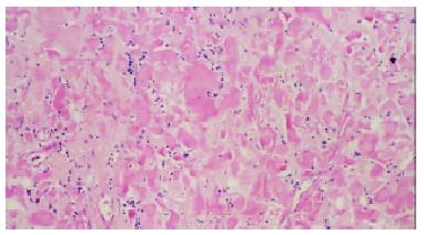
Figure 1 Stained with hematoxylin-eosin, amyloidal deposits in gastric mucosa and submucosa display amorphous, homogeneous, translucent and acidophilic materials under light microscope.
(Magnification × 100).
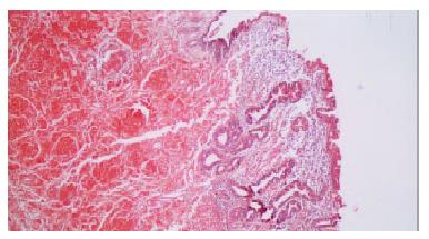
Figure 2 Stained with Congo red, deposition of amyloid could also be observed extending from gastric mucosa to submucosal layer.
(Magnification × 50).
- Citation: Wu D, Lou JY, Chen J, Fei L, Liu GJ, Shi XY, Lin HT. A case report of localized gastric amyloidosis. World J Gastroenterol 2003; 9(11): 2632-2634
- URL: https://www.wjgnet.com/1007-9327/full/v9/i11/2632.htm
- DOI: https://dx.doi.org/10.3748/wjg.v9.i11.2632


