Copyright
©The Author(s) 2003.
World J Gastroenterol. Nov 15, 2003; 9(11): 2523-2527
Published online Nov 15, 2003. doi: 10.3748/wjg.v9.i11.2523
Published online Nov 15, 2003. doi: 10.3748/wjg.v9.i11.2523
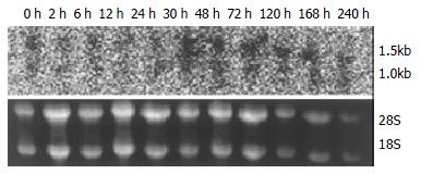
Figure 1 Northern blot of p28GANK mRNA in rat regenerating liver tissues showed two transcripts of 1.
5 kb and 1.0 kb. Expression level of p28GANK mRNA increased 2 h after PH, and reached the peak level at 72 h. More significant variation was found for 1.5 kb transcript.
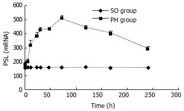
Figure 2 No significant difference of the expression of p28GANK mRNA was seen in the SO group.
In the PH group, the expres-sion increased in 1.5 kb transcript 2 h after PH. The peak time was at 72 h, but gradually decreased after 72 h.
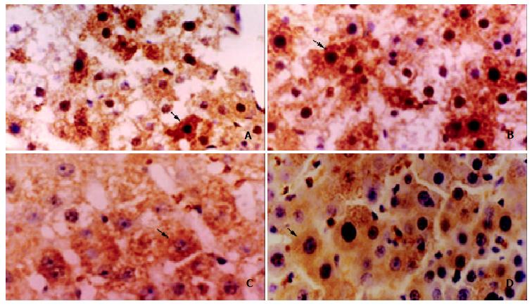
Figure 3 The expression of p28GANK protein in regenerating liver tissues was examined by immunohistochemistry.
A: Local brown expression of p28GANK in the cytoplasm and nucleus of regenerating hepatocyte near central region 24 h after PH. IHC × 400; B: Diffuse brown expression of p28GANK in the cytoplasm and nucleus of regenerating hepatocyte 48 h after PH. IHC × 400; C: Regional light yellow expression of p28GANK in cytoplasm of regenerating hepatocyte 72 h after PH. IHC × 400; D: Dispersed light yellow expression of p28GANK in cytoplasm of regenerating hepatocyte 120 h after PH. IHC × 400.
- Citation: Qin JM, Fu XY, Li SJ, Liu SQ, Zeng JZ, Qiu XH, Wu MC, Wang HY. Gene and protein expressions of p28GANK in rat with liver regeneration. World J Gastroenterol 2003; 9(11): 2523-2527
- URL: https://www.wjgnet.com/1007-9327/full/v9/i11/2523.htm
- DOI: https://dx.doi.org/10.3748/wjg.v9.i11.2523


