Copyright
©The Author(s) 2003.
World J Gastroenterol. Nov 15, 2003; 9(11): 2455-2459
Published online Nov 15, 2003. doi: 10.3748/wjg.v9.i11.2455
Published online Nov 15, 2003. doi: 10.3748/wjg.v9.i11.2455
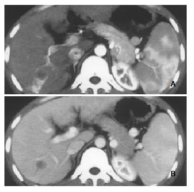
Figure 1 Nodular pattern of HCC with mild and peripheral HAVS.
A: Transient patchy enhancement lateral to HCC foci at late hepatic arterial phase; B: becoming isoattenuation at portal vein phase.
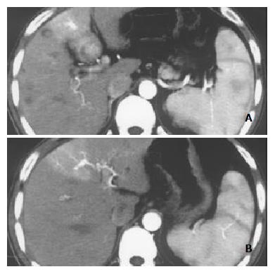
Figure 2 Nodular pattern of HCC accompanied by mild and peripheral HAVS.
Stronger opacification of the third order portal vein branches than that of main portal trunk at late hepatic arterial phase with transient wedge-shaped enhancement lateral to HCC foci (A, B).
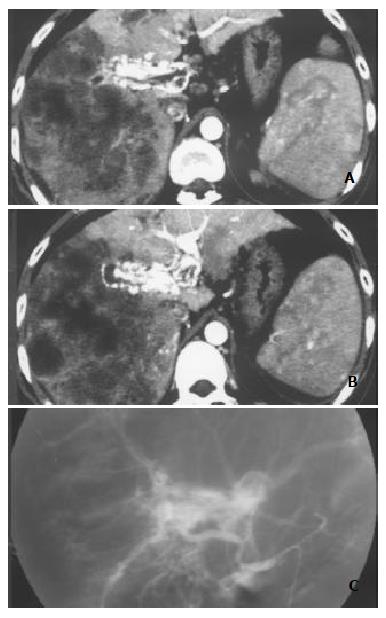
Figure 3 Massive and nodular pattern of HCC associated with severe and central HAVS.
Earlier enhancement and stronger opacification of main portal trunk and the left and right first order branches with thromboses in them were shown. Enhancement degree of HCC foci was decreased, and enhancement degree of liver parenchyma without HCC foci was increased with heterogeneous density (A and B). DSA finding of the same patient (C).
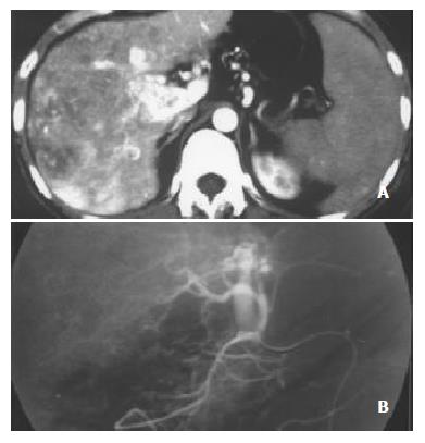
Figure 4 Massive pattern of HCC complicated with severe, central and slight, peripheral HAVS.
Earlier enhancement and stronger opacification of main portal trunk with small thromboses in it were seen, with patchy enhancement internal and lateral to HCC foci. A: Enhancement degrees of HCC foci and spleen were decreased, enhancement degree of liver parenchyma without HCC foci was increased with heterogeneous density; B: DSA finding of the same patient.
- Citation: Luo MY, Shan H, Jiang ZB, Li LF, Huang HQ. Study on hepatocelluar carcinoma-associated hepatic arteriovenous shunt using multidetector CT. World J Gastroenterol 2003; 9(11): 2455-2459
- URL: https://www.wjgnet.com/1007-9327/full/v9/i11/2455.htm
- DOI: https://dx.doi.org/10.3748/wjg.v9.i11.2455


