Copyright
©The Author(s) 2003.
World J Gastroenterol. Jan 15, 2003; 9(1): 69-72
Published online Jan 15, 2003. doi: 10.3748/wjg.v9.i1.69
Published online Jan 15, 2003. doi: 10.3748/wjg.v9.i1.69
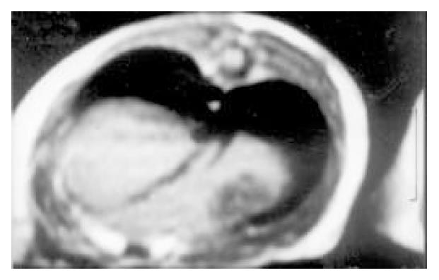
Figure 1 MR imaging showed low signal on T1WI in implanted hepatoma of rat
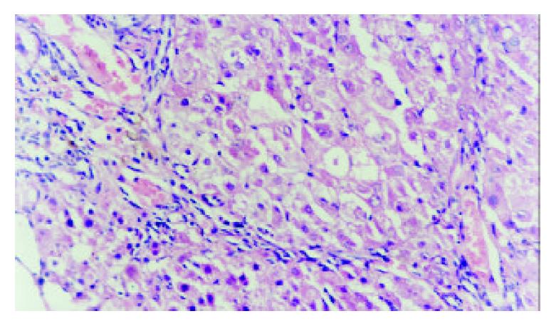
Figure 2 Distribution of microscopy (× 200) in implanted liver cancer that abnormal type and arrangement of cancer cell.
There were abundant vascular proliferation and lymphocyte infiltration growth around tumor
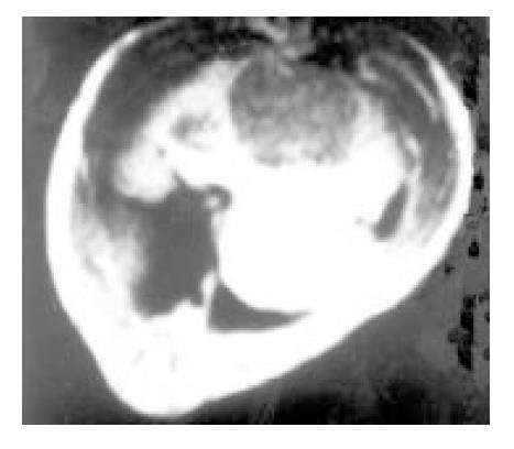
Figure 3 Homogeneous slight hypointensity on T1WI was found in poly-nodules of tumor
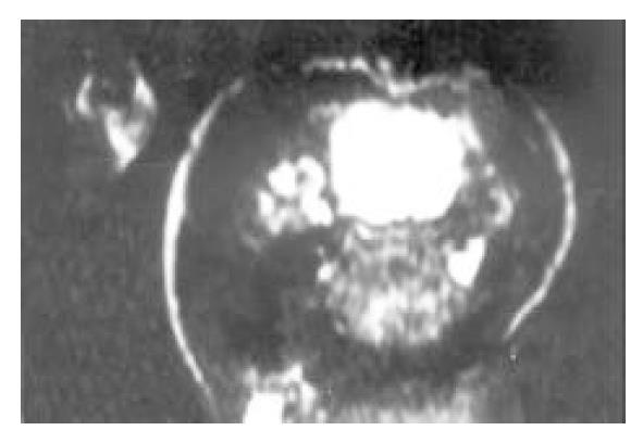
Figure 4 Inhomogeneous slight hyperintensity was indicated on T2WI in different shape, but some of them were showed normal signal or more contrast enhancement in interval
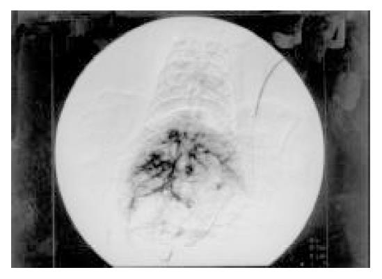
Figure 5 The disorder of distribution of internal hepato-vascularity was found and the angio-clump showed different size in tumor in DSA
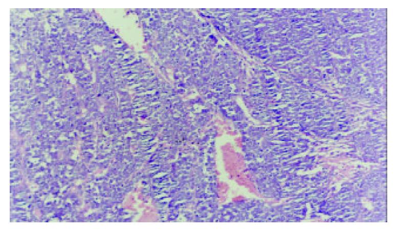
Figure 6 Distribution of microscopy (× 100) in induced liver cancer that tumor and liver tissue was in interval.
There was plenty and dilation of vessel
- Citation: Yang JH, You TG, Li N, Qian QJ, Wang P, Yan ZL, Wu MC. Relationship between the imaging features and pathologic alteration in hepatoma of rats. World J Gastroenterol 2003; 9(1): 69-72
- URL: https://www.wjgnet.com/1007-9327/full/v9/i1/69.htm
- DOI: https://dx.doi.org/10.3748/wjg.v9.i1.69


