Copyright
©The Author(s) 2002.
World J Gastroenterol. Oct 15, 2002; 8(5): 937-942
Published online Oct 15, 2002. doi: 10.3748/wjg.v8.i5.937
Published online Oct 15, 2002. doi: 10.3748/wjg.v8.i5.937
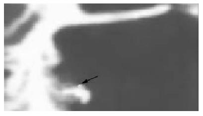
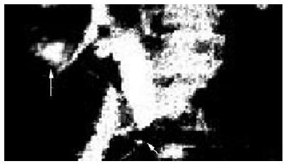
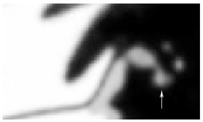
Figure 1 ERCP showed a bile leak from the fossae of gallbladder (↑).
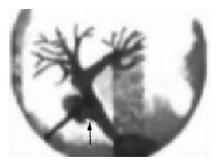
Figure 2 Cholangiography through T tube showed a leak from common bile duct (↑).
Figure 3 ERCP showed a leak from the fistulous tract of T tube (↑) and stones at distal duct (Δ).
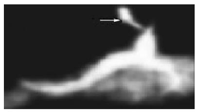
Figure 4 ERCP showed a leak from right hepatic duct (Δ).
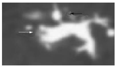
Figure 5 MRCP showed stricture at distal duct (↑) and liquid collection in the right subhepatic region (Δ).
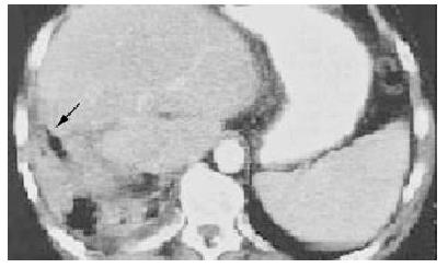
Figure 6 CT showed bile accumulating in the cut surface of right liver (↑).
Figure 7 The distal duct stones was removed with choledochoscope (Δ).
- Citation: Chen XP, Peng SY, Peng CH, Liu YB, Shi LB, Jiang XC, Shen HW, Xu YL, Fang SB, Rui J, Xia XH, Zhao GH. A ten-year study on non-surgical treatment of postoperative bile leakage. World J Gastroenterol 2002; 8(5): 937-942
- URL: https://www.wjgnet.com/1007-9327/full/v8/i5/937.htm
- DOI: https://dx.doi.org/10.3748/wjg.v8.i5.937


