Copyright
©The Author(s) 2002.
World J Gastroenterol. Oct 15, 2002; 8(5): 832-836
Published online Oct 15, 2002. doi: 10.3748/wjg.v8.i5.832
Published online Oct 15, 2002. doi: 10.3748/wjg.v8.i5.832
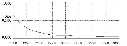
Figure 1 The UV spectrum adsorption curve of GBSP from 200 nm to 400 nm
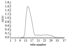
Figure 2 The profile of Sepharose 4B gel filtration chromatography
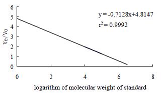
Figure 3 Standard linear calibration curve of Sephadex G-200 chromatography
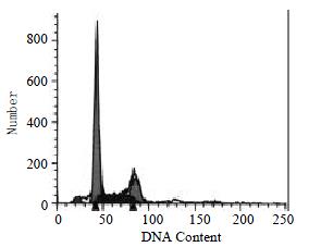
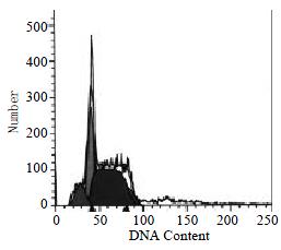
Figure 4 In the absence of GBSP treatment, the apoptosis ratio of SMMC-7721 cells was 3.
84% ± 0.55%, the cells debris was 0.46% ± 0.12% and the percentage of G2-M cells was 17.01% ± 1.28%.
Figure 5 In the presence of GBSP treatment, the apoptosis ratio of SMMC-7721 cells was 9.
13% ± 1.48%, the cells debris was 0.06% ± 0.06%, and the percentage of G2-M cells was 11.77% ± 1.50%.
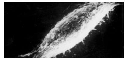
Figure 6 The majority of SMMC-7721 cells were of shuttle shape without GBSP treatment (× 2500)
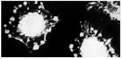
Figure 7 There were more spherical SMMC-7721 cells after GBSP treatment (× 2200).
These cells shrunk and apoptosis bodies were formed on and around them.
-
Citation: Chen Q, Yang GW, An LG. Apoptosis of hepatoma cells SMMC-7721 induced by
Ginkgo biloba seed polysaccharide. World J Gastroenterol 2002; 8(5): 832-836 - URL: https://www.wjgnet.com/1007-9327/full/v8/i5/832.htm
- DOI: https://dx.doi.org/10.3748/wjg.v8.i5.832


