Copyright
©The Author(s) 2002.
World J Gastroenterol. Aug 15, 2002; 8(4): 707-711
Published online Aug 15, 2002. doi: 10.3748/wjg.v8.i4.707
Published online Aug 15, 2002. doi: 10.3748/wjg.v8.i4.707
Figure 1 p38 MAPK activities in the course of ischemia and reperfusion.
Livers were perfused through the portal vein with KH-buffer and snap frozen at 30 min, after 24 h of ischemic storage in UW solution (4 °C), and after 45 and 120 min of reperfusion. Deep-frozen organs were lysed and assayed for p38 MAPK activity as described under "Materials and Methods" Determination of density light units was performed by phosphorimaging and values for the respective time points are expressed as ration vs values for pre-ischemic (30 min) organs. Bars show means ± SEM of three independent perfusion experiments. aP < 0.05 and bP < 0.01: statistically different from pre-ischemic 30 min controls.
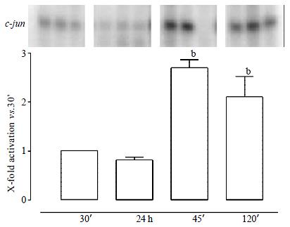
Figure 2 JNK activities in the course of ischemia and reperfusion.
Isolated perfused rat livers were snap frozen at 30 min, after 24 h of ischemic storage in UW solution (4 °C), and after 45 and 120 min of reperfusion. Deep-frozen organs were lysed and assayed for MAPK activity as described under "Materials and Methods" Determination of density light units was performed by phosphorimaging and values for the respective time points are expressed as ration vs values for pre-ischemic (30 min) organs. Bars show means ± SEM of three independent perfusion experiments. bP < 0.01: statistically different from pre-ischemic 30 min controls.
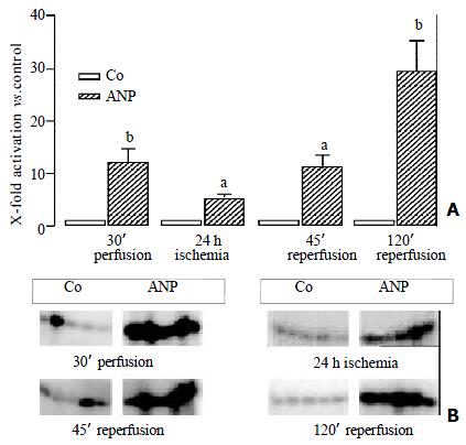
Figure 3 ANP increases the activity of p38 MAPK in the course of ischemia and reperfusion.
Livers perfused with KH-buffer in the presence or absence of ANP (200 nM) underwent 24 h of cold ischemic storage and were snap frozen at the respective time points. p38 MAPK activity was determined as described under "Materials and Methods" Density light units were determined by phosphorimaging and values for ANP-treated organs were divided by mean values for control organs at the respective time points. Panel A: Bars show means ± SEM of five independent perfusion experiments with aP < 0.05 and bP < 0.01: statistically different from controls at the respective time point. Panel B: Data show representative autoradiograms.
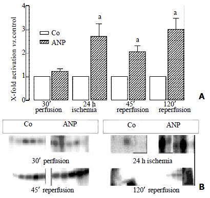
Figure 4 ANP and JNK activity.
Isolated perfused livers were either left untreated or preconditioned with ANP (200 nM) for 20 min. After 24 h of ischemia and up to 120 min of reperfusion JNK activity was determined as described under "Materials and Methods" Determination of density light units was performed by phosphorimaging and values for ANP-treated organs were divided by mean values for control organs at the respective time points. Panel A: Bars show means ± SEM of five independent perfusion experiments with aP < 0.05: statistically different from controls at the respective time point. Panel B: Data show representative autoradiograms.
- Citation: Kiemer AK, Kulhanek-Heinze S, Gerwig T, Gerbes AL, Vollmar AM. Stimulation of p38 MAPK by hormal preconditioning with atrial natriuretic peptide. World J Gastroenterol 2002; 8(4): 707-711
- URL: https://www.wjgnet.com/1007-9327/full/v8/i4/707.htm
- DOI: https://dx.doi.org/10.3748/wjg.v8.i4.707


