Copyright
©The Author(s) 2002.
World J Gastroenterol. Aug 15, 2002; 8(4): 638-643
Published online Aug 15, 2002. doi: 10.3748/wjg.v8.i4.638
Published online Aug 15, 2002. doi: 10.3748/wjg.v8.i4.638
Figure 1 Northern blot of p28/gankyrin in a series of human normal tissues, hepatoma cell lines, HCC and non-tumor liver tissues.
(A) p28/gankyrin expression in ten different normal adult human tissues. (B) p28/gankyrin expression in five hepatoma cell lines and NIH/3T3 cell. (C) p28/gankyrin expression in HCC and para-carcinoma liver tissues. L indicates the para-carcinoma liver tissues, and K indicates the HCC tissues. (D) Comparison of p28/gankyrin expression in normal human liver tissues, liver cirrhosis and HCC tissues. Each bottom panel showed equal amount of total RNA loading as indicated in 28 s and 18 s rRNA.
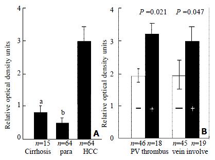
Figure 2 Densitometric analysis.
The ratio of the optical density of p28/gankyrin mRNA to the corresponding 18 S rRNA signals was calculated and expressed as means ± SD. (A) Comparison of p28/gankyrin expression in human liver cirrhosis, para-carcinoma liver tissues and HCCs. aThe expression in HCCs was significantly higher than that in liver cirrhosis (P < 0.05). bThe expression in HCCs was significantly higher than that in para-carcinoma liver tissues (P < 0.01). (B) Clinicopathological significance of p28/gankyrin expression in HCCs. p28/gankyrin mRNA levels were significantly higher (P = 0.021) in portal vein thrombus group than in non-thrombus group, higher (P = 0.047) in microscopic hepatic vein involvement group than in non-hepatic vein involvement group.
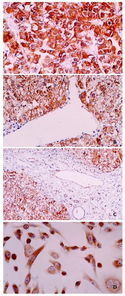
Figure 3 Immunohistochemical staining of P28/Gankyrin in HCC, para-carcinoma liver tissue and SK-Hep1 cell line.
(A) p28/gankyrin was moderately to intensely present in the cytoplasm of most hepatocytes in HCC tissue, and occasionally present in the nucleus of hepatocytes; (B) In contrast, p28/gankyrin was weakly present in the cytoplasm of hepatocytes in para-carcinoma liver tissue; (C) p28/gankyrin protein signals were specifically present in the cytoplasm of hepatocytes, while absent in bile duct cells, blood vein endometrial cells and other interstitial cells; (D) In SK-Hep1 cells, p28/gankyrin was mostly located in cytoplasm and occasionally in the nucleus.
-
Citation: Fu XY, Wang HY, Tan L, Liu SQ, Cao HF, Wu MC. Overexpression of
p 28/gankyrin in human hepatocellular carcinoma and its clinical significance. World J Gastroenterol 2002; 8(4): 638-643 - URL: https://www.wjgnet.com/1007-9327/full/v8/i4/638.htm
- DOI: https://dx.doi.org/10.3748/wjg.v8.i4.638


