Copyright
©The Author(s) 2001.
World J Gastroenterol. Dec 15, 2001; 7(6): 805-810
Published online Dec 15, 2001. doi: 10.3748/wjg.v7.i6.805
Published online Dec 15, 2001. doi: 10.3748/wjg.v7.i6.805
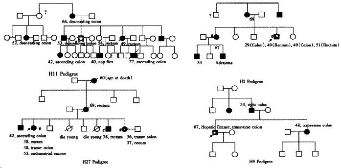
Figure 1 Four typical Chinese HNPCC pedigrees.
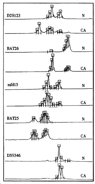
Figure 2 MSI status of H2 proband.
Microsatellite analysis of H2 proband with five microsatellite markers, MSI in 4/5 loci.
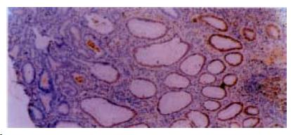
Figure 3 Immunohistochemical staining.
No hMSH2 protein expression in adenoma and carcinoma areas of H11 proband tumor section, infiltrating lymphocytes as well as normal colonic crypt epithelium next to the tumor showed nuclear staining of the hMSH2 protein. × 100
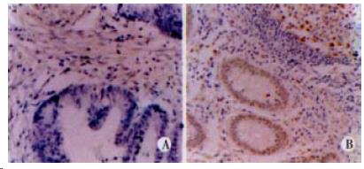
Figure 4 Immunohistochemical staining.
A: No hMLH1 protein expression in carcinoma area of H9 proband tumor section. × 400 B: Infiltrating lymphocytes as well as normal colonic crypt epithelium next to the tumor showed nuclear staining of the hMLH1 protein. × 200
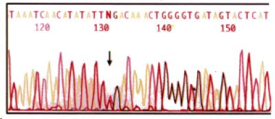
Figure 5 C-T transition (CGA-TGA) at codon 680 in ex on 13 of hMSH2 gene in H2 proband, resulting in a stop codon.
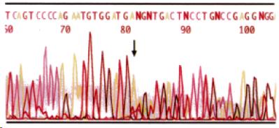
Figure 6 A 24 bp deletion at codon 305 in exon 11 of hMLH1 gene in H9 proband.
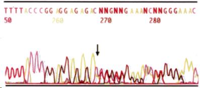
Figure 7 A 1 bp insertion at codon 206 in exon 3 of hMSH2 gene in H27 proband, resulting in a stop codon 73 bp downstream of the mutation.
- Citation: Cai Q, Sun MH, Lu HF, Zhang TM, Mo SJ, Xu Y, Cai SJ, Zhu XZ, Shi DR. Clinicopathological and molecular genetic analysis of 4 typical Chinese HNPCC families. World J Gastroenterol 2001; 7(6): 805-810
- URL: https://www.wjgnet.com/1007-9327/full/v7/i6/805.htm
- DOI: https://dx.doi.org/10.3748/wjg.v7.i6.805


