Copyright
©The Author(s) 2001.
World J Gastroenterol. Oct 15, 2001; 7(5): 672-677
Published online Oct 15, 2001. doi: 10.3748/wjg.v7.i5.672
Published online Oct 15, 2001. doi: 10.3748/wjg.v7.i5.672
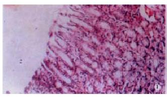
Figure 1 Grade 1 damage in gastric corpus and antrum.
LM × 330. In grade 1 damage, surface mucous cells were damaged.
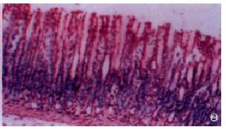
Figure 2 Grade 1 damage in gastric corpus and antrum.
LM × 330. In grade 1 damage, surface mucous cells were damaged.
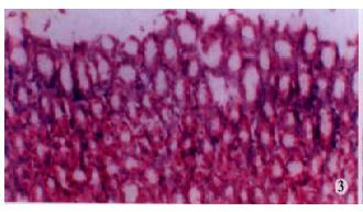
Figure 3 Grade 2 damage in gastric corpus and antrum.
LM × 330. In grade 2 damage, the cells kining the gastric pits were also disrupted.
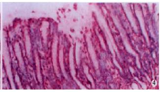
Figure 4 Grade 2 damage in gastric corpus and antrum.
LM × 330. In grade 2 damage, the cells kining the gastric pits were also disrupted.
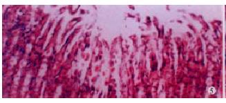
Figure 5 Grade 3 damage in gastric corpus and antrum.
LM × 330. In grade 3 damage, cell destruction extended into the gastric glands.
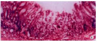
Figure 6 Grade 3 damage in gastric corpus and antrum.
LM × 330. In grade 3 damage, cell destruction extended into the gastric glands.
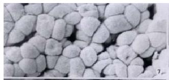
Figure 7 Grade 1 damage in gastric corpus.
SEM × 1500.
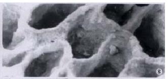
Figure 8 Grade 2 damage in gastric corpus.
SEM × 1500.
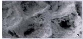
Figure 9 Grade 3 damage in gastric corpus.
SEM × 1500.
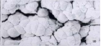
Figure 10 Grade 1 damage in gastric antrum.
SEM × 1500. In grade 1 damage, surface cells were of irregular shape, and gaps between indiv idual cells were present.
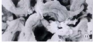
Figure 11 Grade 2 damage in gastric antrum.
SEM × 1500. In grade 2 damage, the basal lamina was exposed, but still showed continuity. Wide openings to the gastric pits were visible.
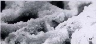
Figure 12 Grade 3 damage in gastric antrum.
SEM × 1500. In grade 3 damage, most of the basal lamina was disrupted, Regular surface cells were no longer present.
- Citation: Zhang LH, Yao CB, Li HQ. Effects of extract F of red-rooted Salvia on mucosal lesions of gastric corpus and antrum induced by hemorrhagic shock-reperfusion in rats. World J Gastroenterol 2001; 7(5): 672-677
- URL: https://www.wjgnet.com/1007-9327/full/v7/i5/672.htm
- DOI: https://dx.doi.org/10.3748/wjg.v7.i5.672


