Copyright
©The Author(s) 2001.
World J Gastroenterol. Feb 15, 2001; 7(1): 74-79
Published online Feb 15, 2001. doi: 10.3748/wjg.v7.i1.74
Published online Feb 15, 2001. doi: 10.3748/wjg.v7.i1.74
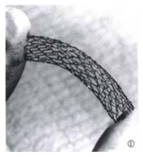
Figure 1 The Cordis balloon-expandable stent, which is constructed as a sinusoidal wave pattern by one stainless steel wire, with high rigidity.
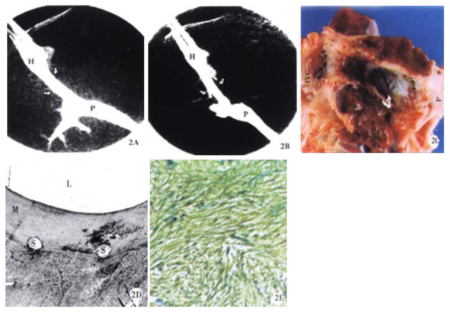
Figure 2 a.
The portal venography obtained immediately shows that the TIPS shunt (arrows) is well created between the hepatic vein (H) and the portal vein (P). b. The shunt remains patent in the portography taken immediately before the euthanasia at 2 weeks after stent placement. c. The gross examination of the specimen shows that the surface of the stent (straight arrows) is covered by a smooth and thin layer endothelia-like tissue (curved arrows) which does not extend to the ends of the stent at the IVC and the portal vein (P). d. Low magnification photomicrography of the TIPS reaction shows a mild myofibroblastic cell proliferation (M) under stent meshes (S) (Modified Giemsa and basic Fuchsin stain, × 3.12). e. The myofibroblastic cells are primarily composed of smooth muscle cells. (Anti-SMCα actin stain, × 125).
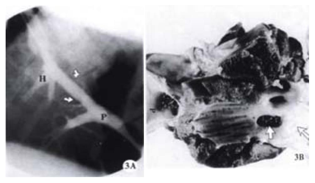
Figure 3 a.
An occluded TIPS shunt from a pig using a Cordis stent. The immediate portal venography after TIPS shows a patent TIPS shunt (arrows). b. The occluded shunt was confirmed by the portography obtained immediately before the euthanasia in two weeks after the stent placement. The gross examination of the specimen demonstrates that the hepatic end of the stent wedges into the liver parenchyma and the proximal end of the shunt is totally occluded by the healed hepatic vein (arrows), while the portal end (P) of the stent keeps patent.
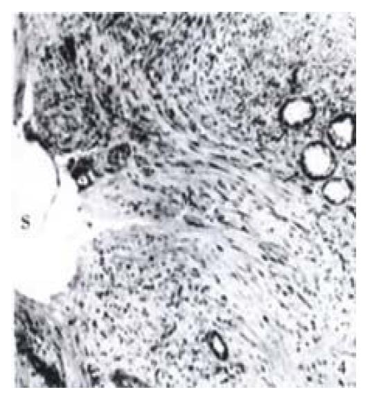
Figure 4 Photomicrography from an occluded TIPS shunt using a Wallstent shows that a massive pseudointimal proliferative tissue under the stent (S), which primarily composed of myofibroblastic cells (M).
Neo-bile duct proliferation within the proliferation is also noted (arrows). Modified Giemsa and basic Fuchsin stain, × 125
- Citation: Teng GJ, Bettmann MA, Hoopes PJ, Yang L. Comparison of a new stent and Wallstent for transjugular intrahepatic portosystemic shunt in a porcine model. World J Gastroenterol 2001; 7(1): 74-79
- URL: https://www.wjgnet.com/1007-9327/full/v7/i1/74.htm
- DOI: https://dx.doi.org/10.3748/wjg.v7.i1.74


