Copyright
©The Author(s) 2000.
World J Gastroenterol. Dec 15, 2000; 6(6): 920-924
Published online Dec 15, 2000. doi: 10.3748/wjg.v6.i6.920
Published online Dec 15, 2000. doi: 10.3748/wjg.v6.i6.920
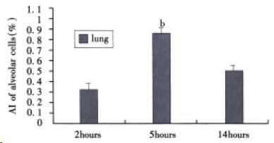
Figure 1 AI of alveolar epithelial cells in sodium taurocholate-induced pancreatitis-associated lung injury.
AHNP was induced by retrograde injections into the pancreatic ducts of 5% sodium taurocholate (0.1 mL/100 g body wt) in rats. Results shown are the MEAN ± SEM for four or more animals in each group. Asterisks indicates bP < 0.01 when the AI of alveolar epithelial cells at 5 h after induction of AHNP compared with 2 and 14 h after induction of AHNP.
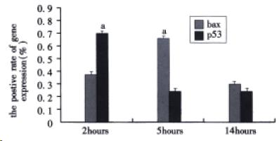
Figure 2 The positive rate of bax and p53 protein in alveolar epithelial cells after sodium taurocholate-induced pancreatitis-associated lung injury.
AHNP was induced by retrograde injections into the pancreatic ducts of 5% sodium taurocholate (0.1 mL/100 g body wt) in rats. Results shown were the MEAN ± SEM for four or more animals in each group. Asterisks indicates aP < 0.05 when the positive rate of bax protein in alveolar epithelial cells at 5 h after induction of AHNP compared with 2 and 14 h and the positive rate of p53 protein in alveolar epithelial cells at 2 h after induction of AHNP compared with 5 and 14 h.
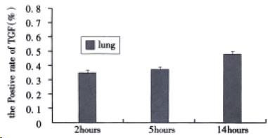
Figure 3 The positive rate of TGFβ1 in alveolar epithelial cells after sodium taurocholate-induced pancreatitis-associated lung injury.
AHNP was induced by retrograde injections into the pancreatic ducts of 5% sodium taurocholate (0.1 mL/100 g body wt) in rats. Results shown were the MEAN ± SEM for four or more animals in each group. Therewas no significant difference among the positive rate of TGFβ1 at 2, 5 and 14 h after induction of AHNP.
- Citation: Yuan YZ, Gong ZH, Lou KX, Tu SP, Zhai ZK, Xu JY. Involvement of apoptosis of alveolar epithelial cells in acute pancreatitis-associated lung injury. World J Gastroenterol 2000; 6(6): 920-924
- URL: https://www.wjgnet.com/1007-9327/full/v6/i6/920.htm
- DOI: https://dx.doi.org/10.3748/wjg.v6.i6.920


