Copyright
©The Author(s) 2000.
World J Gastroenterol. Dec 15, 2000; 6(6): 861-865
Published online Dec 15, 2000. doi: 10.3748/wjg.v6.i6.861
Published online Dec 15, 2000. doi: 10.3748/wjg.v6.i6.861
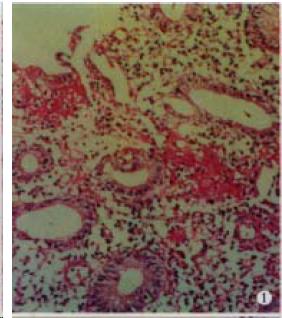
Figure 1 The colonic mucosa shows hyperemia, edema and inflammatory cell infiltration.
The goblet cells in the body of gland decrease remarkably, which are composed of columnar cells.
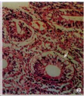
Figure 2 The lesion is the same as above.
The arrowhead shows an infranuclear vacuole.
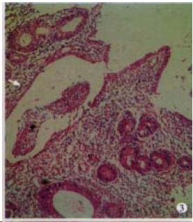
Figure 3 The lesion is the same as above.
The arrowhead shows a crypt abscess. H.E. × 100
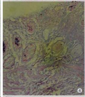
Figure 4 The majority of acid mucin in the body of gland disappear.
AB-PAS staining × 100
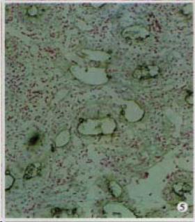
Figure 5 The majority of mucin sulfate in the body of gland disappears.
HID-AB staining × 100
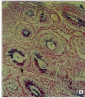
Figure 6 The mucin in the body of gland decre ases.
The amount of neutral mucin relatively increases (arrowhead). AB-PAS staining × 100
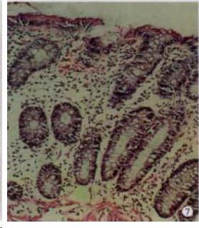
Figure 7 The colonic mucosa shows mild hyperemia, with less mucosal inflammatory cell infiltration.
The goblet cells in the body of gland show no reduction. H.E. × 100
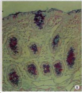
Figure 8 The reduction of acid mucin is not obvious.
AB-PAS staining × 100
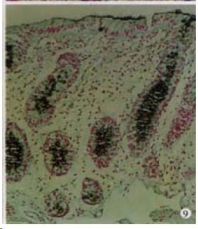
Figure 9 No reduction of mucin sulfate occurs.
HID-AB staining × 100
- Citation: Wu HG, Zhou LB, Shi DR, Liu SM, Liu HR, Zhang BM, Chen HP, Zhang LS. Morphological study on colonic pathology in ulcerative colitis treated by moxibustion. World J Gastroenterol 2000; 6(6): 861-865
- URL: https://www.wjgnet.com/1007-9327/full/v6/i6/861.htm
- DOI: https://dx.doi.org/10.3748/wjg.v6.i6.861


