Copyright
©The Author(s) 2000.
World J Gastroenterol. Dec 15, 2000; 6(6): 842-847
Published online Dec 15, 2000. doi: 10.3748/wjg.v6.i6.842
Published online Dec 15, 2000. doi: 10.3748/wjg.v6.i6.842
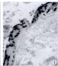
Figure 1 Distribution of CAL-IRE cells (↑) in esophageal epithelium of M.
albus. × 66.( Note: ★:goblet cell; L: gastrointestinal lumen; Is: pancreatic island; P: pancreas
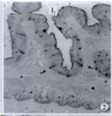
Figure 2 SOM-IRE cells (↑) in intestinal epithelium of T.
nilotica. × 33
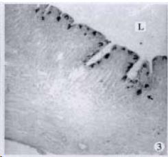
Figure 3 GAS-IRE cells (↑) in gastric epithelium of C.
argus. × 33
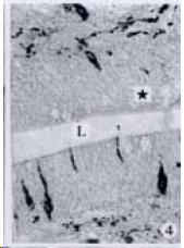
Figure 4 5-HT-IRE cells (↑) in intestinal epithelium of C.
argus. × 100
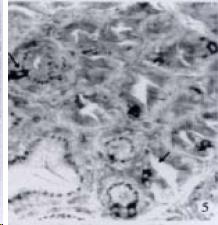
Figure 5 SOM-IRE cells (↑) in gastric gland of P.
fulvidraco. × 152
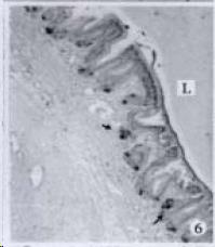
Figure 6 SOM-IRE cells (↑) in pyloric gland of P.
fulv idraco. × 33
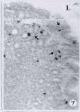
Figure 7 SOM-IRE cells (↑) in cardiac epithelium and gland of S.
chuatsi. × 66
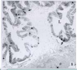
Figure 8 5-HT-IRE cells in intestinal epithelium (↑) and intermuscular nerve plexus (△) of S.
chuatsi. × 33
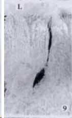
Figure 9 Shape of 5-HT-IRE cells (↑) in gastric epithelium and glands of M.
salmoides. × 200
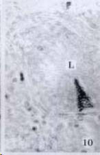
Figure 10 Shape of 5-HT-IRE cells (↑) in gastric epithelium and glands of M.
salmoides. × 200
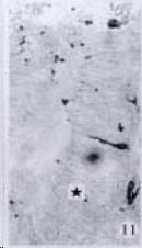
Figure 11 5-HT-IRE cells (↑) in intestinal epithelium of M.
salmoides. × 100
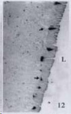
Figure 12 5-HT-IRE cells (↑) in gastric epithelium of S.
asotus. × 33
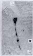
Figure 13 Shape of 5-HT-IRE cells (↑) in intestinal epithelium of S.
asotus. × 200
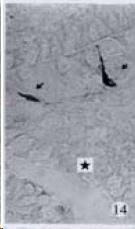
Figure 14 5-HT-IRE cells (↑) in intestinal epithelium of C.
brachypomum. × 200
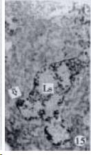
Figure 15 GLU-IRE cells (△) in pancreatic islands of S.
chuatsi. × 33
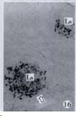
Figure 16 SOM-IRE cells (△) in pancreatic islands of S.
asotus. × 33
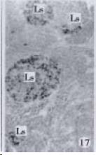
Figure 17 INS-IRE cells (△) in pancreatic islands of S.
chuatsi. × 33
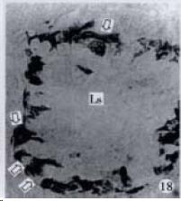
Figure 18 Shape of GLU-IRE cells (△) in pancreatic islands S.
asotus. × 268
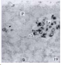
Figure 19 GH-IRE cells in pancreatic islands (△) and pancreas (↑) of S.
asotus. × 200
- Citation: Pan QS, Fang ZP, Huang FJ. Identification, localization and morphology of APUD cells in gastroenteropancreatic system of stomach-containing teleosts. World J Gastroenterol 2000; 6(6): 842-847
- URL: https://www.wjgnet.com/1007-9327/full/v6/i6/842.htm
- DOI: https://dx.doi.org/10.3748/wjg.v6.i6.842


