Copyright
©The Author(s) 2000.
World J Gastroenterol. Dec 15, 2000; 6(6): 819-823
Published online Dec 15, 2000. doi: 10.3748/wjg.v6.i6.819
Published online Dec 15, 2000. doi: 10.3748/wjg.v6.i6.819
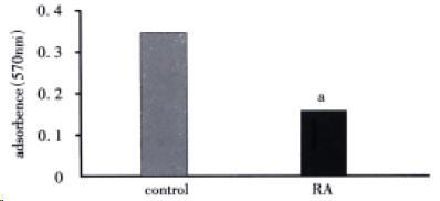
Figure 1 RA inhibited HSC proliferation.
TGF -β1-stimulated rat HSC were cultured and treated with or without 1 nmol/L RA for 48 h, followed by MTT assay as described in MATERIALS AND METHODS. aP < 0.05 vs control.
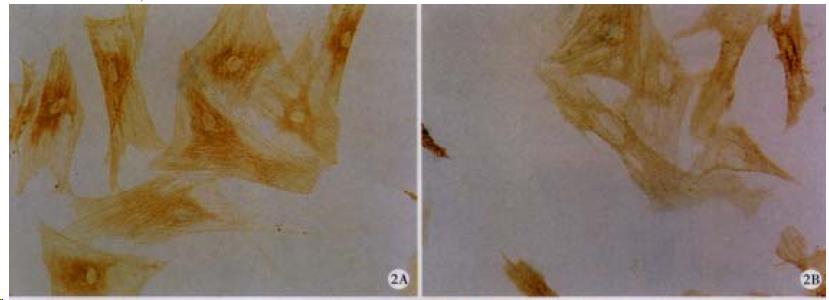
Figure 2 RA decreased the protein level of α -SMA.
TGF-β1-stimulated HSC were treated with (A) as descr ibed in Figure 1, and then immunocytochemistry was performed to detect α-SMA protein. ABC × 200 (B) or without RA
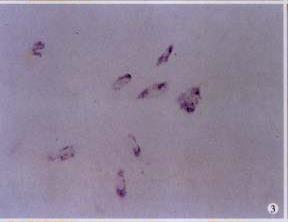
Figure 3 Expression of RAR-β2 in RA-treated HSC.
In situ hybridization with DIG-labeled RAR-β2 cDNA probe was used to determine RAR-β2 mRNA expression in HSC. NBT/BCIP × 200
Figure 4 Expression of CKI protein.
Immunocyt ochemical study was performed to detect CKI, i.e. p16 (A); p21 (B); ABC × 200 (C), expression in control (A) or RA-treated HSC
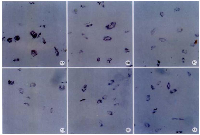
Figure 5 mRNA expression of CKI.
mRNA of p16 (A), p21, (B), (C) or p27, (D) was determined with ISH in control (E) or RA-treated HSC (F). NBT/BCIP × 200
- Citation: Huang GC, Zhang JS, Zhang YE. Effects of retinoic acid on proliferation, phenotype and expression of cyclin-dependent kinase inhibitors in TGF-β1-stimulated rat hepatic stellate cells. World J Gastroenterol 2000; 6(6): 819-823
- URL: https://www.wjgnet.com/1007-9327/full/v6/i6/819.htm
- DOI: https://dx.doi.org/10.3748/wjg.v6.i6.819


