Copyright
©The Author(s) 2000.
World J Gastroenterol. Oct 15, 2000; 6(5): 725-729
Published online Oct 15, 2000. doi: 10.3748/wjg.v6.i5.725
Published online Oct 15, 2000. doi: 10.3748/wjg.v6.i5.725
Figure 1 Structure of bicistronic retroviral vector PGCEN/β-gal.
Arrow below vector indicates initiated site of transcription.
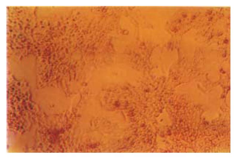
Figure 2 Morphology of hepatocytes in culture.
× 100 Hepatocytes became polygonal epitheliumlike structure. The majority of cells were mononucleated; Some cells were bi-or multi-nucleated. The membranes were visible. Hepatocytes became confluent at 4 d postplating.
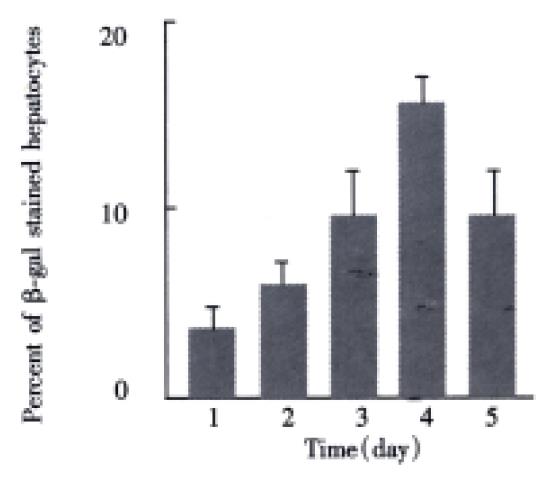
Figure 3 Transduction efficiency of hepatocytes by retroviral vector.
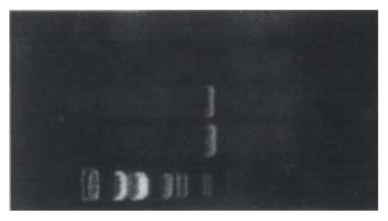
Figure 4 PCR detection of NeoR in primary rat hepatocytes transduced by PGCEN/β-gal.
A: Size markers (Lambda DNA/EcoR + Hind III Marker) B: Positive template (MN45Li cell lines modified by NeoR gene) C: Primary rat hepatocytes transduced with retroviral vector PGCEN/β-gal D: Nontransduced primary rat hepatocytes E: Water control
- Citation: Xie Q, Liao D, Zhou XQ, Qian SB, Cheng SS. Transduction of primary rat hepatocytes with bicistronic retroviral vector. World J Gastroenterol 2000; 6(5): 725-729
- URL: https://www.wjgnet.com/1007-9327/full/v6/i5/725.htm
- DOI: https://dx.doi.org/10.3748/wjg.v6.i5.725


