Copyright
©The Author(s) 2000.
World J Gastroenterol. Oct 15, 2000; 6(5): 681-687
Published online Oct 15, 2000. doi: 10.3748/wjg.v6.i5.681
Published online Oct 15, 2000. doi: 10.3748/wjg.v6.i5.681
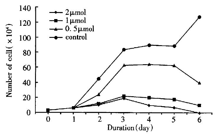
Figure 1 The effect of As2O3 on cell growth of BEL-7402.
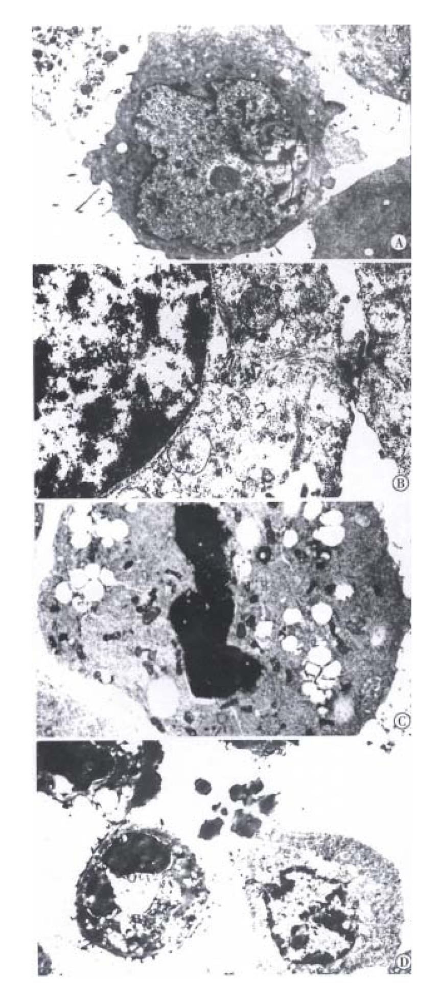
Figure 2 Transmission electronmicroscopic observation of ultrastructural changes in human hepatoma cells treated with 0, 0.
5, 1, 2 μmol/L As2O3 for 96 h. The nucleocytoplasmic ratio enlanged with indentation of nuclei in BEL-7402 cells of 0 μmol/L As2O3 group (A). The nucleocytoplasmic ratio become smaller, nuclei appear round, but with well-differentiated organelles in the cytoplasm in 0.5 μmol/L As2O3 group (B). The intact cell membrane, nuclear condensation and apoptoti c body formation in 1, 2 μmol/L As2O3 group (C, D).
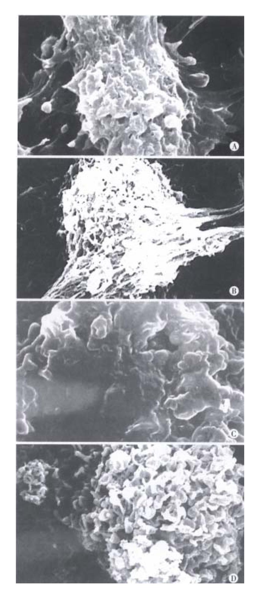
Figure 3 Scanning electronmicroscopic observation of changes in human hepatoma cell membranes treated with 0, 0.
5, 1, 2 μmol/L As2O3 for 96 h. There were abundant processes and micro villi on the surface of BEL-7402 cells in 0 μmol/L As2O3 group (A). The processes and micro villi was much less in 0.5 μmol/L As2O3 group (B). The processes and micro villi were lost when treated with 1, 2 μmol/L As2O3 (C, D).
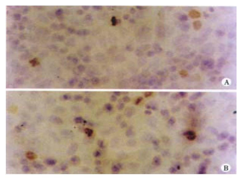
Figure 4 Bcl-2 expressions in human hepatoma cells.
0 μmol/L As2O3 group (A). 0.5 μmol/L As2O3 group (B).
-
Citation: Xu HY, Yang YL, Gao YY, Wu QL, Gao GQ. Effect of arsenic trioxide on human hepatoma cell line BEL-7402 cultured
in vitro . World J Gastroenterol 2000; 6(5): 681-687 - URL: https://www.wjgnet.com/1007-9327/full/v6/i5/681.htm
- DOI: https://dx.doi.org/10.3748/wjg.v6.i5.681


