Copyright
©The Author(s) 2000.
World J Gastroenterol. Oct 15, 2000; 6(5): 676-680
Published online Oct 15, 2000. doi: 10.3748/wjg.v6.i5.676
Published online Oct 15, 2000. doi: 10.3748/wjg.v6.i5.676
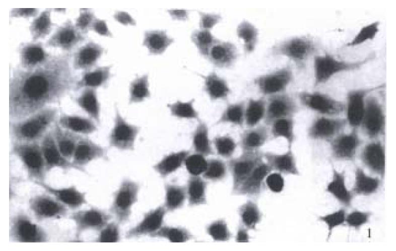
Figure 1 BGC-823 cells.
× 300
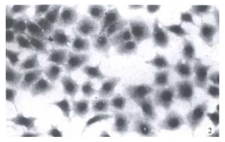
Figure 2 BGC-823 cells treated with tachyplesin.
× 300
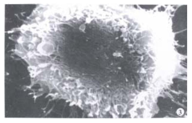
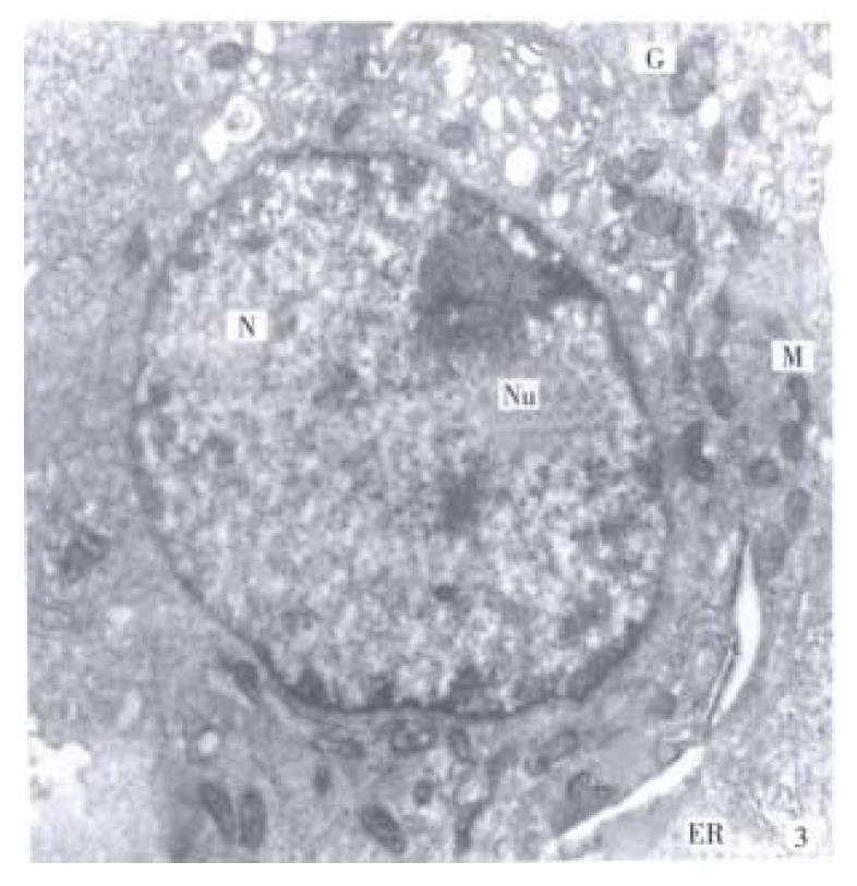
Figure 3 There are abundant microvilli on spherical cell surface and many filopodia emerge at the edge of BGC-823 cells.
× 6000
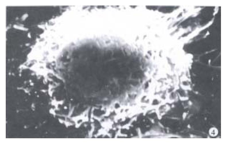
Figure 4 The microvilli on spherical cell surface disappear mainly in the cells after tachyplesin treatment.
× 6000
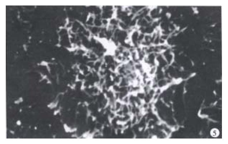
Figure 5 The microvilli are plentiful on flat and spread cell surface and many filopodia appear at the edge of BGC-823 cells.
× 4800
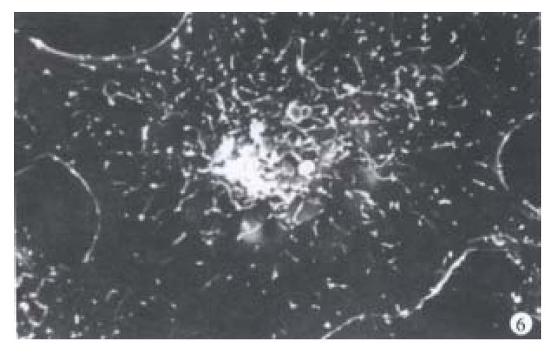
Figure 6 On the cells treated with tachyplsin the microvilli are rare, shrink and shortened on flat and spread cell surface and large lame llipodia emerge at cell edge.
× 4200
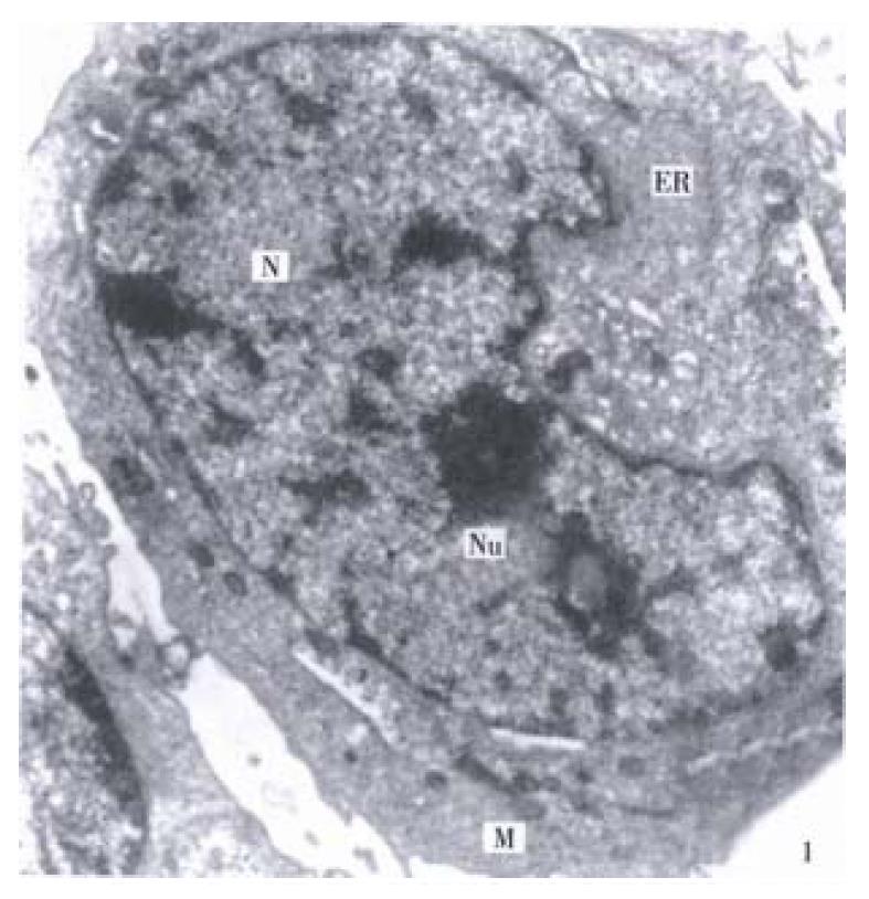
Figure 7 The nucleo-cytoplasm ratio is large, the shape of nucleus (N) is irregular and only a few organelle in cytoplasm of BGC-823 cells (Nu nucleolus, M mitochordria, ER endoplasmic reticulum).
× 9600
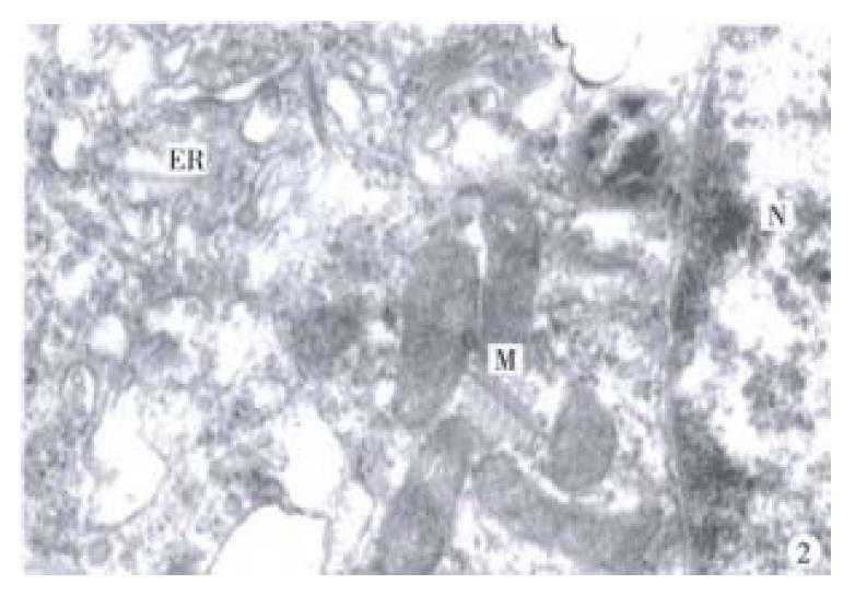
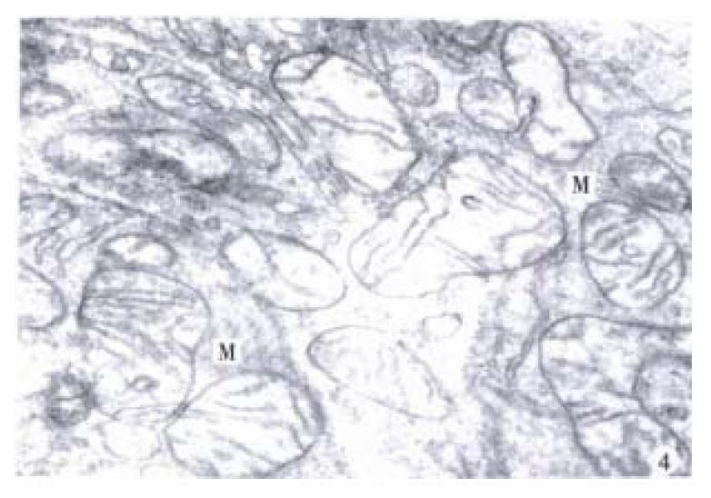
Figure 8 The shape and structure of mitochondria (M) is non-typical and endoplasmic reticulum (ER) decrease in BGC-823 cells.
× 40500
Figure 9 The cellular volume enlarge, the nucleo-cytoplasm ratio decreases and the organelle increase in the cells treated with tachyplesin.
× 10720
Figure 10 The shape and structure of mitochondria (M) are unanimity relatively and their cristae increase and arrange rather regularly in the cells treated with tachyplsin.
× 21000
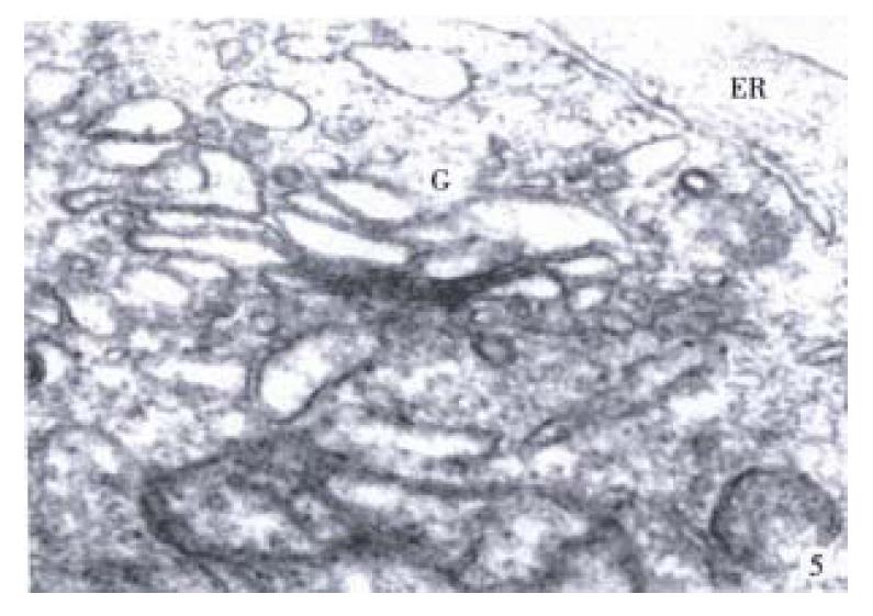
Figure 11 The structure of Golgi complex (G) appear typical and well-developed in the cells treated with tachyplsin.
× 48000
- Citation: Li QF, Ou-Yang GL, Li CY, Hong SG. Effects of tachyplesin on the morphology and ultrastructure of human gastric carcinoma cell line BGC-823. World J Gastroenterol 2000; 6(5): 676-680
- URL: https://www.wjgnet.com/1007-9327/full/v6/i5/676.htm
- DOI: https://dx.doi.org/10.3748/wjg.v6.i5.676


