Copyright
©The Author(s) 2000.
World J Gastroenterol. Jun 15, 2000; 6(3): 458-460
Published online Jun 15, 2000. doi: 10.3748/wjg.v6.i3.458
Published online Jun 15, 2000. doi: 10.3748/wjg.v6.i3.458
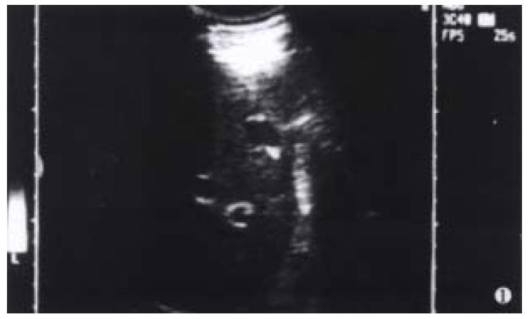
Figure 1 A nodule appeared as well-defined, hypoechoic and hypovascular irregular solid mass by ultrasonography.
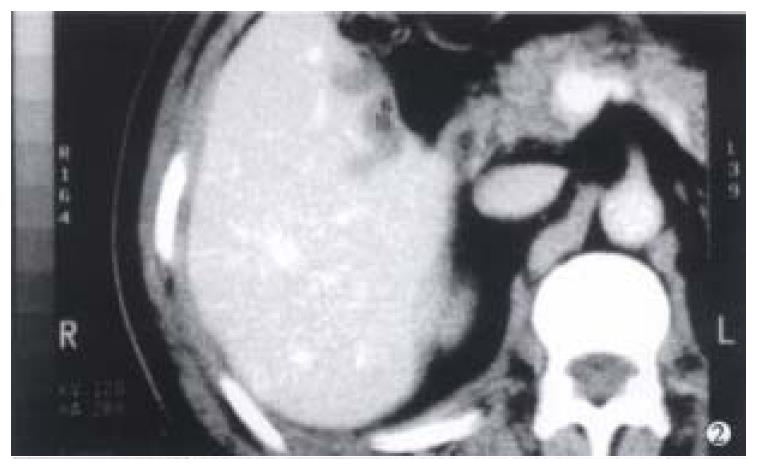
Figure 2 Two nodules of low density without early enhancement detected on CT scans.
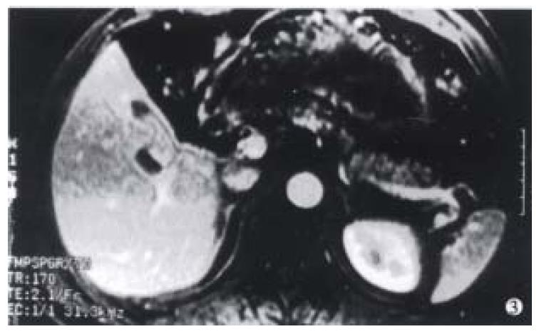
Figure 3 Two nodules of low signal intensity on T1WI and iso-signal intensity on T2WI by MRI.
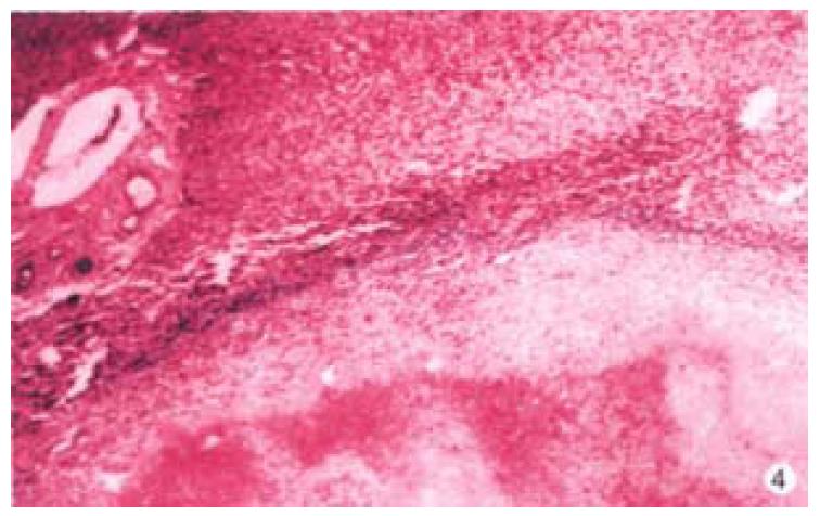
Figure 4 Solitary necrotic nodules of the liver.
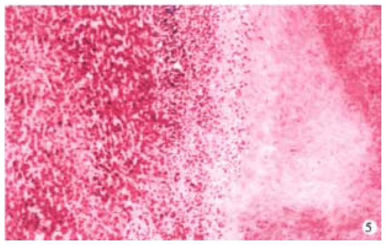
HE × 50
Figure 5 Central necrotic core enclosed by a hyalinised fibrotic capsule.
HE × 100
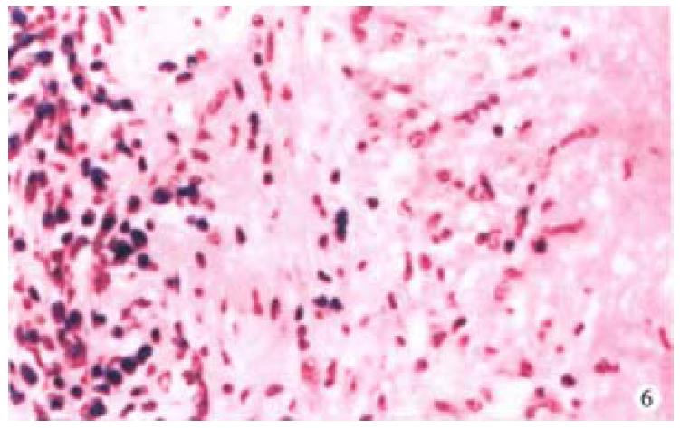
Figure 6 Hyalinised fibrotic capsule infiltrated with eosinophils and lymphocytes.
HE × 200
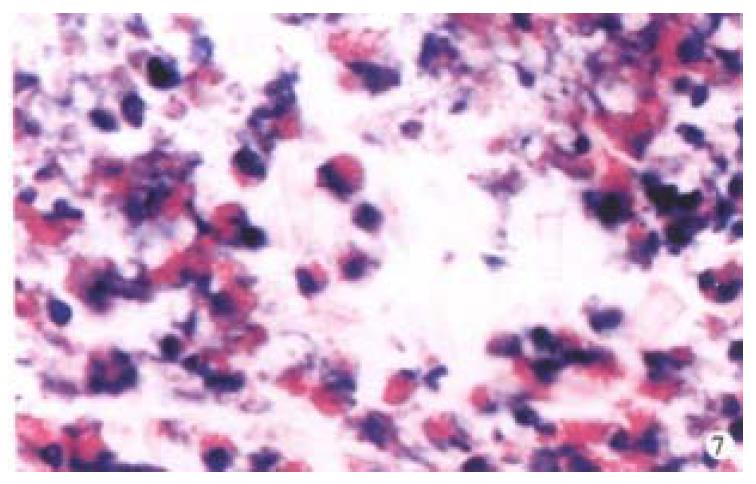
Figure 7 Charcot Leyden's crystals and eosinophils in the necrotic center.
HE × 400
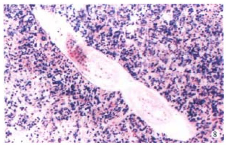
Figure 8 One degenerated larva in the center of necrotic nodule.
HE × 100
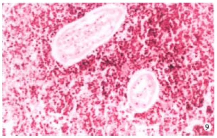
Figure 9 Two pieces of the degenerated larva in the necrotic center of the nodule.
HE × 200
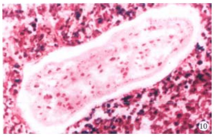
Figure 10 Necrotic cells around the larva.
HE × 200
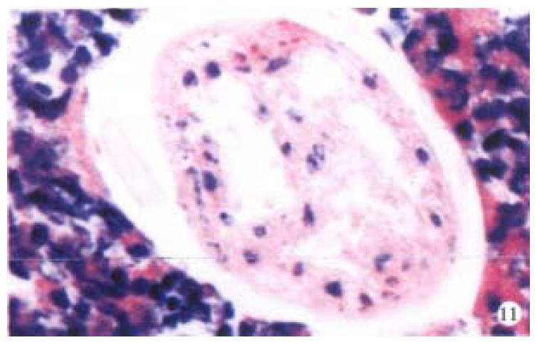
Figure 11 Degenerated larva with Charcot Leyden's crystals.
HE × 400
- Citation: Ji XL, Shen MS, Yin T. Liver inflammatory pseudotumor or parasitic granuloma? World J Gastroenterol 2000; 6(3): 458-460
- URL: https://www.wjgnet.com/1007-9327/full/v6/i3/458.htm
- DOI: https://dx.doi.org/10.3748/wjg.v6.i3.458


