Copyright
©The Author(s) 2000.
World J Gastroenterol. Jun 15, 2000; 6(3): 353-355
Published online Jun 15, 2000. doi: 10.3748/wjg.v6.i3.353
Published online Jun 15, 2000. doi: 10.3748/wjg.v6.i3.353
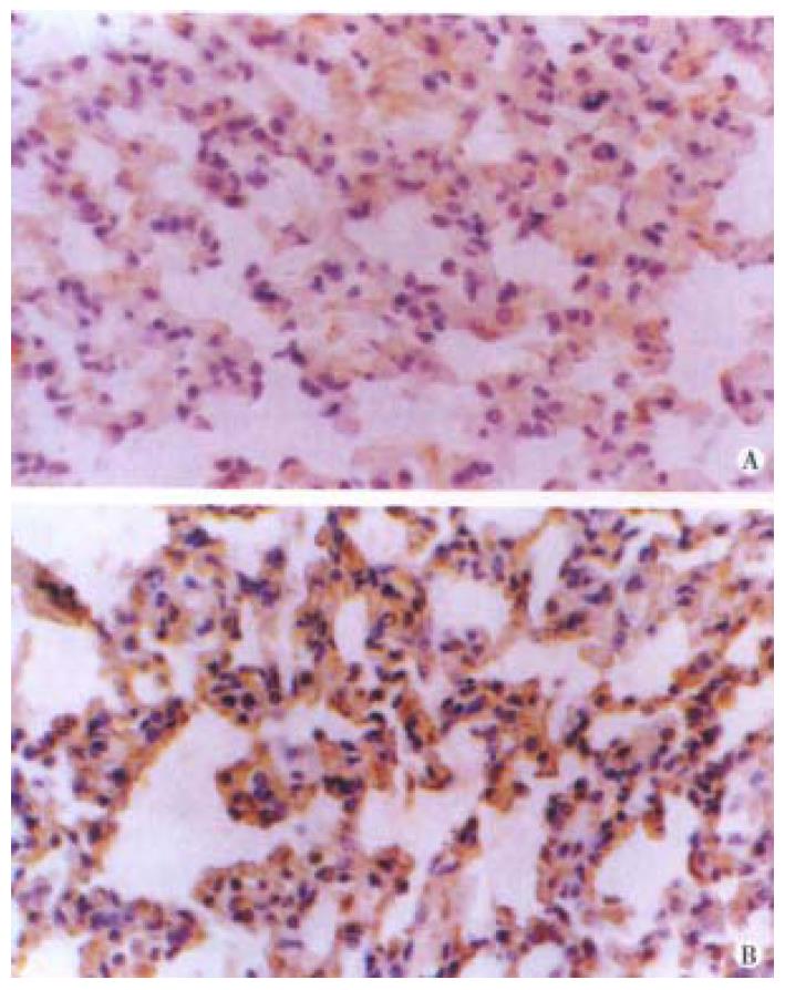
Figure 1 The expressions of bFGF (A) and TGF-β (B) in normal lung.
The weakly positive signal could be found in alveolar epithelial cells and microvascular endothelial cells. SP stain × 400
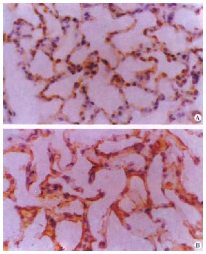
Figure 2 The expressions of bFGF (A) and TGF-β (B) in damaged lung following ischemia (45 min) and reperfusion (6 h).
Deep staining of both growth factors could be found in alveolar epithelial cells and microvascular endothelial cells. SP stain × 400
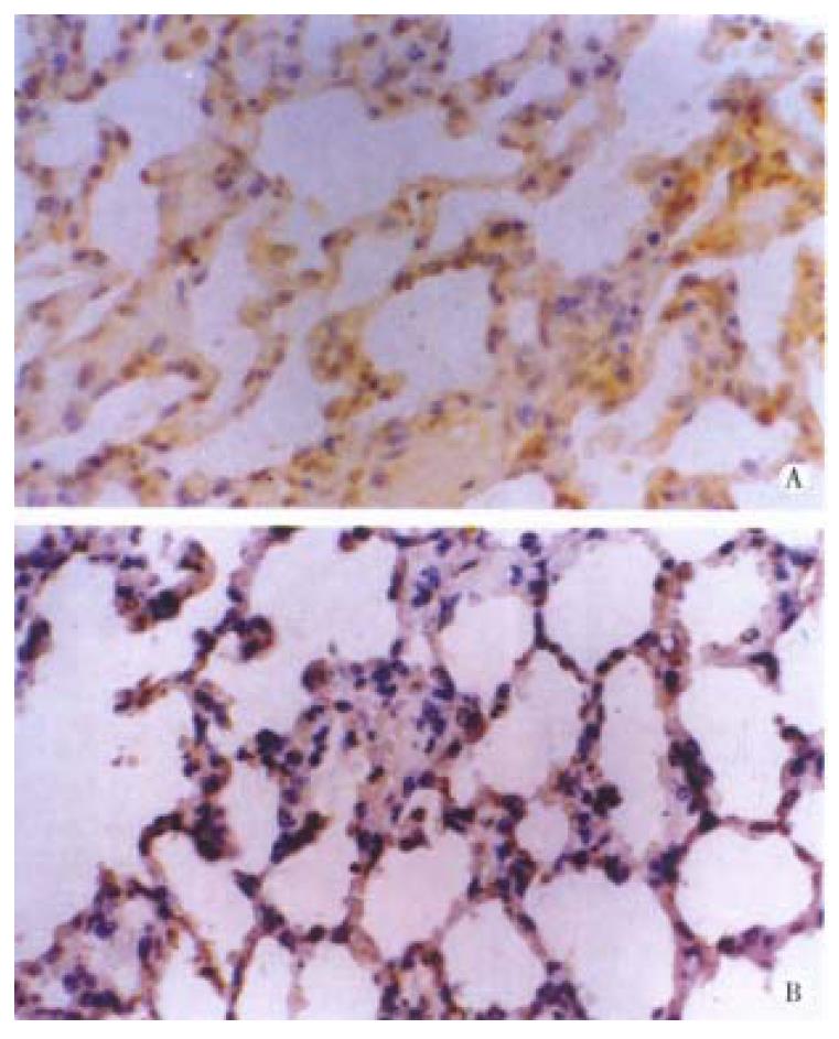
Figure 3 The expressions of bFGF (A) and TGF-β (B) in damaged lung following ischemia (45 min) and reperfusion (24 h).
The staining of both growth factors could be found in alveolar epithelial cells and microvascular endothelial cells and the expression returned to normal levels. SP stain × 400
- Citation: Fu XB, Yang YH, Sun TZ, Gu XM, Jiang LX, Sun XQ, Sheng ZY. Effect of intestinal ischemia-reperfusion on expressions of endogenous basic fibroblast growth factor and transforming growth factor β in lung and its relation with lung repair. World J Gastroenterol 2000; 6(3): 353-355
- URL: https://www.wjgnet.com/1007-9327/full/v6/i3/353.htm
- DOI: https://dx.doi.org/10.3748/wjg.v6.i3.353


