Copyright
©The Author(s) 2000.
World J Gastroenterol. Apr 15, 2000; 6(2): 263-265
Published online Apr 15, 2000. doi: 10.3748/wjg.v6.i2.263
Published online Apr 15, 2000. doi: 10.3748/wjg.v6.i2.263
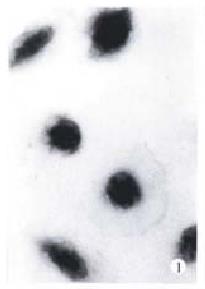
Figure 1 Control BEL-7402 cells, W-G staining.
× 350
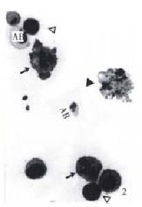
Figure 2 BEL-7402 cells treated with 10 mg/L NCTD for 24 h.
(↑) indicates multipolar distribution of chromosomes in postmitotic cells. (▲) nuclear fragments in apoptotic cells, (-) indicates con densed cells, (AB) apoptotic bodes containing one or two nuclear fragments. W-G staining, × 350
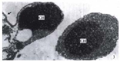
Figure 3 Ultrastructural features of BEL-7402 cells treated with 10 mg/L NCTD for 24 h.
Endoplasmic reticulum (ER) dilated in the apoptotic cell (↑), the apoptotic bodies characterized by compaction of nuclear chromatin and condensation of cytoplasm. × 14000
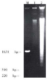
Figure 4 Agarose gel electrophoresis of DNA extracted from BEL-7402 cells.
M: DNA marker; 1: control group; 2: group treated with 10 mg/L NCTD for 24 h.
Figure 5 Western blotting analysis of Bcl-2 protein in BEL-7402 cells.
1: control; 2: treated with 10 mg/L NCTD for 24 h; 3: treated with 10 mg/L NCTD for 48 h; 4: treated with 10 mg/L NCTD for 72 h.
- Citation: Sun ZX, Ma QW, Zhao TD, Wei YL, Wang GS, Li JS. Apoptosis induced by norcantharidin in human tumor cells. World J Gastroenterol 2000; 6(2): 263-265
- URL: https://www.wjgnet.com/1007-9327/full/v6/i2/263.htm
- DOI: https://dx.doi.org/10.3748/wjg.v6.i2.263


