Copyright
©The Author(s) 2000.
World J Gastroenterol. Apr 15, 2000; 6(2): 220-222
Published online Apr 15, 2000. doi: 10.3748/wjg.v6.i2.220
Published online Apr 15, 2000. doi: 10.3748/wjg.v6.i2.220
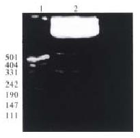
Figure 1 Incomplete digestion of plasmid pQ534 by restriction endonucleases Sma I and Pst I.
Lane 1: DNA molecular weight marker, lane 2: pQ534 digested with Pst I first, and then digested incompletely.
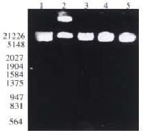
Figure 2 Identification of the recombinant plasmid pTM3-Q534 with restriction endonucleases.
Lane 1: 500 ng Hind-III/EcoR I DNA marker; lane 2: pTM3 digested with Sma-I and Pst I; lanes 3 and 4: pTM3-Q534 digested with Eco-R I and Pst I; lane 5: pTM3-Q534 digested with Eco-R I.
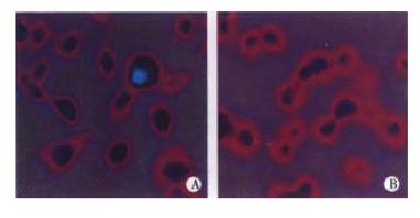
Figure 3 Indirect immunofluorescence staining of transfected HepG2 cells.
A: A part of the HepG2 cells infected with vvT-7.3 and transfected with pTM3-Q534 showing intracellular immunofluorescence; B: In control, no staining can be seen in the HepG2 cells infected with vvT-7.3 and transfected with pTM3.
- Citation: Jiang RL, Lu QS, Luo KX. Cloning and expression of core gene cDNA of Chinese hepatitis C virus in cosmid pTM3. World J Gastroenterol 2000; 6(2): 220-222
- URL: https://www.wjgnet.com/1007-9327/full/v6/i2/220.htm
- DOI: https://dx.doi.org/10.3748/wjg.v6.i2.220


