Copyright
©The Author(s) 1999.
World J Gastroenterol. Dec 15, 1999; 5(6): 541-543
Published online Dec 15, 1999. doi: 10.3748/wjg.v5.i6.541
Published online Dec 15, 1999. doi: 10.3748/wjg.v5.i6.541
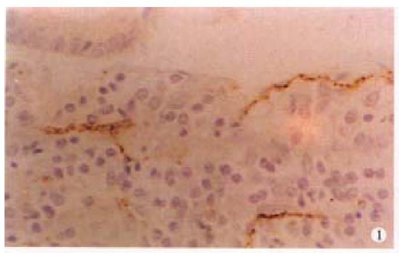
Figure 1 VIP immunoreactive nerve fibers were coarse in mucosa of CD.
ABC method × 400
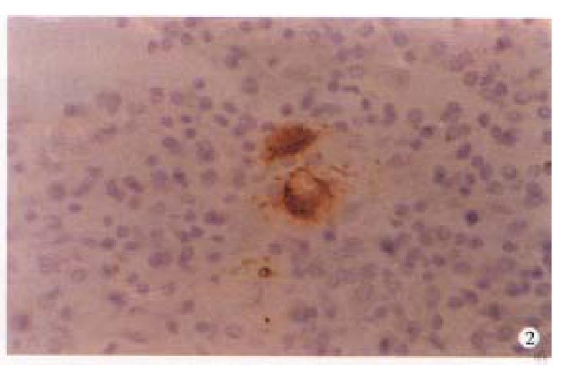
Figure 2 VIP immunoreactive neurons were hypertrophi al in submucosa of CD.
ABC method × 400
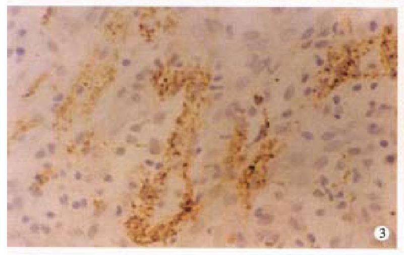
Figure 3 VIP immunoreactive nerve fibers were irregu larly thickened in submucosa of CD.
ABC method × 400
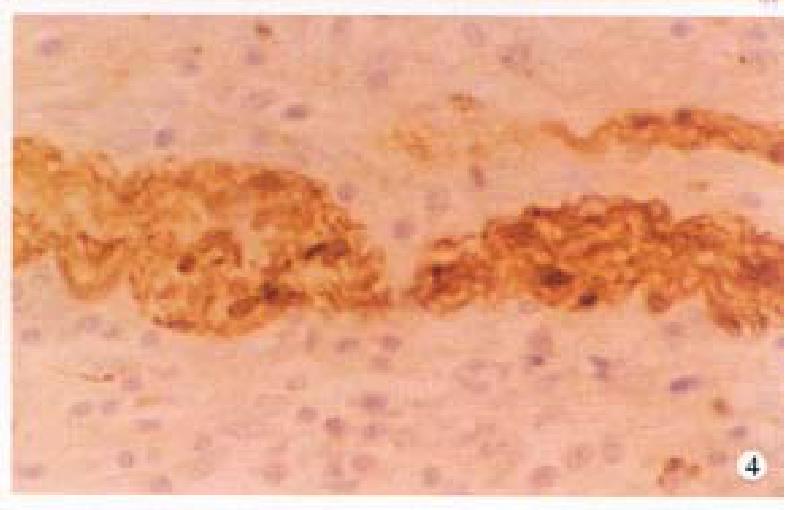
Figure 4 S-100 protein immunoreactive nerve fibers were increased in myenteric plexus of CD.
ABC method × 400
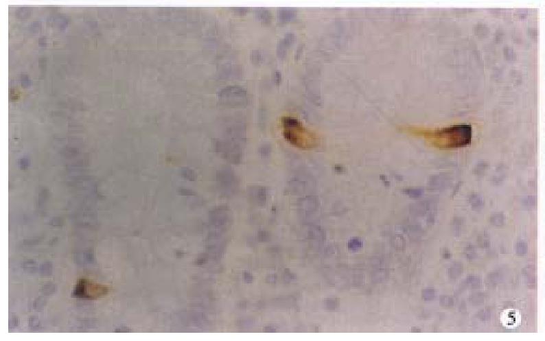
Figure 5 Serotonin immunoreactive cells in mucosa of CD.
ABC method × 400
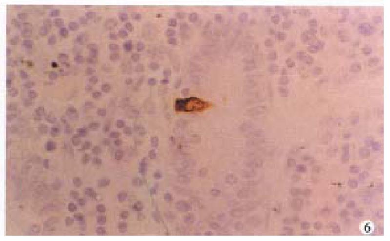
Figure 6 Somatostatin immunoreactive cells in mucosa of CD.
ABC method × 400
- Citation: Lu SJ, Liu YQ, Lin JS, Wu HJ, Sun YH, Tan YB. VIP immunoreactive nerves and somatostatin and serotonin containing cells in Crohn’s disease. World J Gastroenterol 1999; 5(6): 541-543
- URL: https://www.wjgnet.com/1007-9327/full/v5/i6/541.htm
- DOI: https://dx.doi.org/10.3748/wjg.v5.i6.541


