Copyright
©The Author(s) 1999.
World J Gastroenterol. Dec 15, 1999; 5(6): 465-469
Published online Dec 15, 1999. doi: 10.3748/wjg.v5.i6.465
Published online Dec 15, 1999. doi: 10.3748/wjg.v5.i6.465
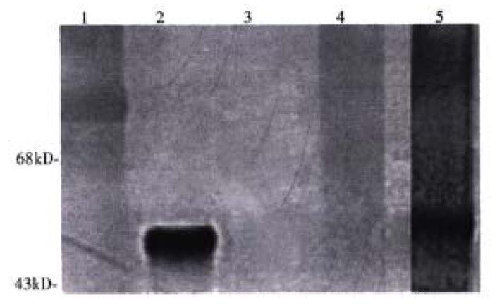
Figure 1 A Western blot of purified conjugates.
One milligram of LLO and the final conjugate either with or without DDT (100 mM) redu ction were run on a 7.5% SDS-PAGE gel. The proteins were transferred to anylon membrane, quenched, and probed with a polyclonal antibody to LLO. Detection of the antigen-antibody complex was determined by exposure to anti-rabbit IgG ho rseradish peroxidase and developed with 3’,3’-diaminobenzidine and hydrogen peroxide as described in Materials and Methods. Molecular weight markers, lane 1; LLO alone, lane 2; ASOR-PL, lane 3; ASOR-PL-LLO-DNA, lane 4; ASOR-PL-LLO-DNA + 100 mM DTT,lane 5.
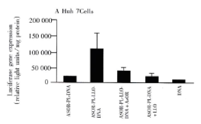
Figure 2 Targeted luciferase gene expression.
Conjugates containing 1μg of CMV luc were added to Huh7 (ASG receptor positive) or SK Hep1 cells (ASG receptor negative) and incubated for 48 h as describ ed in Materials and Methods. Gene expression was measured by luciferase detection using the luciferin substrate, and detection of activity using a luminometer. Luciferase expression results were standardized by measuring protein concentrati ons according to the Bradford assay. Panel A, Huh7 cells: ASOR-PL-DNA, lane 1; ASOR-PL-LLO-DNA, lane 2; ASOR-PL-LLO-DNA+200-fold molar excess of ASOR, lane 3; ASOR-PL-DNA+LLO, lane 4; DNA alone, lane 5. Panel B, SK Hep1 cells: AS OR-PL-DNA, lane 1; ASOR-PL-LLO-DNA, lane 2; ASOR-PL DNA+LLO, lane 3; DNA a lone, lane 4.
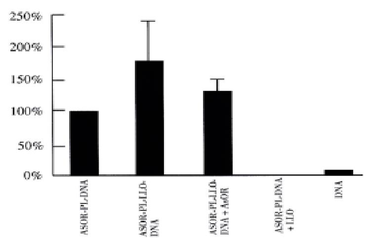
Figure 3 Targeted β-galactosidase gene expression.
Conjugates containing 1μg of β-gal DNA were added to Huh7 (ASG receptor positive) or SK Hepl cells (ASG receptor negative) and incubated for 48 h. Cells incubated with â-gal were washed with PBS pH 74, fixed with 4% paraf ormaldehyde and stained with X-gal as described in Materials and Methods. Cells were observed with light microscopy and positive (blue stained) cells were coun ted. ASOR-PL-DNA, lane 1; ASOR-PL-LLO-DNA, lane 2; ASOR-PL-LLO-DNA+ASOR, lane 3; ASOR-PL-DNA+LLO, lane 4; DNA alone, lane 5.
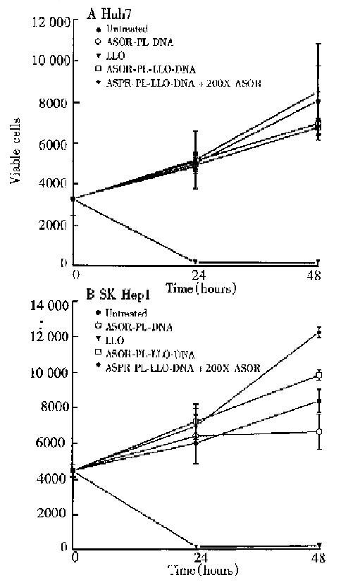
Figure 4 Toxicity of complexes.
Huh7 (ASG receptor p ositive) and SK Hep1 (ASG receptor negative) cells were incubated with ASOR-PL-DNA, free LLO, ASOR-PL-LLO-DNA or ASOR-PL-LLO-DNA in the presence of a 200-fold excess of ASOR as described in Materials and Methods. Cell viability was determined by trypan blue exclusion. Panel A, Huh7 cells; Panel B SK Hep1 cells.
- Citation: Walton CM, Wu CH, Wu GY. A DNA delivery system containing listeriolysin O results in enhanced hepatocyte-directed gene expression. World J Gastroenterol 1999; 5(6): 465-469
- URL: https://www.wjgnet.com/1007-9327/full/v5/i6/465.htm
- DOI: https://dx.doi.org/10.3748/wjg.v5.i6.465


