Copyright
©The Author(s) 1999.
World J Gastroenterol. Apr 15, 1999; 5(2): 167-171
Published online Apr 15, 1999. doi: 10.3748/wjg.v5.i2.167
Published online Apr 15, 1999. doi: 10.3748/wjg.v5.i2.167
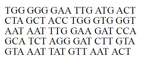
Figure 1 Partial sequence of HBeAg amplified by PCR.
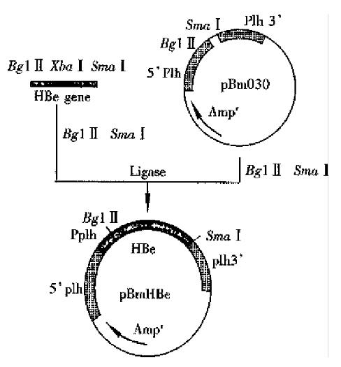
Figure 2 Construction of recombinant vector pBm HBe.
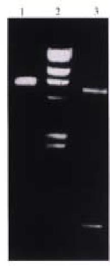
Figure 3 Restriction endonuclease analysis plasmid pBm HBe (Left).
1. p Bm HBe/Bgl II, linerization; 2. λDNA/Hind III marker; 3. p Bm Hbe/Bg-II + Sma 1 I, released 0.54 kb HBeAg gene.
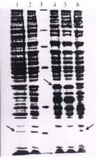
Figure 4 PAG Electrophoresis analysis of HBeAg prote in expressed in Bm N cells(Right).
1 and 2: Bm N cells infected withr Bm-HBe, the arrow shows HBeAg protein, Mr18000; 3: Standard protein molecular weight marker, Mr94000, 17000, 43000, 20000, 17500; 4 and 5: Bm- N cells infected with wt Bm NPV, the arrow shows Mr32000 polyhe dea protein; 6: Culture supernatant, the arrow shows HBeAg protein.
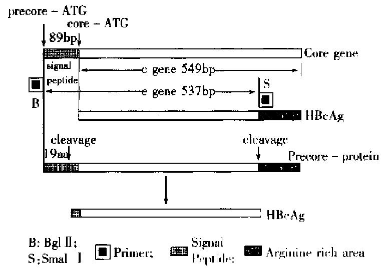
Figure 5 Structure of HBeAg gene and PCR design.
-
Citation: Deng XZ, Diao ZY, He L, Qiao RL, Zhang LY.
HBeAg gene expression with baculovirus vector in silk worm cells. World J Gastroenterol 1999; 5(2): 167-171 - URL: https://www.wjgnet.com/1007-9327/full/v5/i2/167.htm
- DOI: https://dx.doi.org/10.3748/wjg.v5.i2.167


