Copyright
©The Author(s) 1998.
World J Gastroenterol. Jun 15, 1998; 4(3): 214-218
Published online Jun 15, 1998. doi: 10.3748/wjg.v4.i3.214
Published online Jun 15, 1998. doi: 10.3748/wjg.v4.i3.214
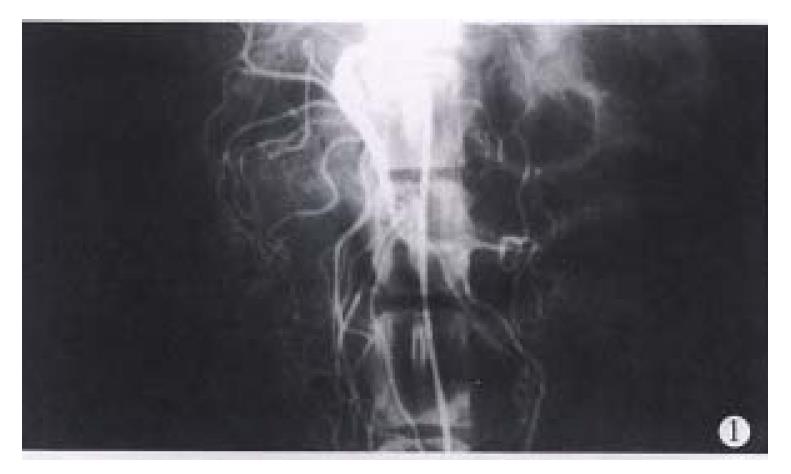
Figure 1 Anterior mesenteric arteriography in a dog.
The point of the catheter is in the stem of the anterior mesenteric artery.
Figure 2 The point entered the right place of the dog’s spleen.
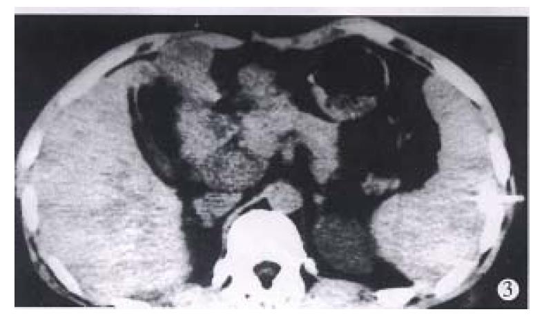
Figure 3 The point entered the right place of the patient’s spleen.
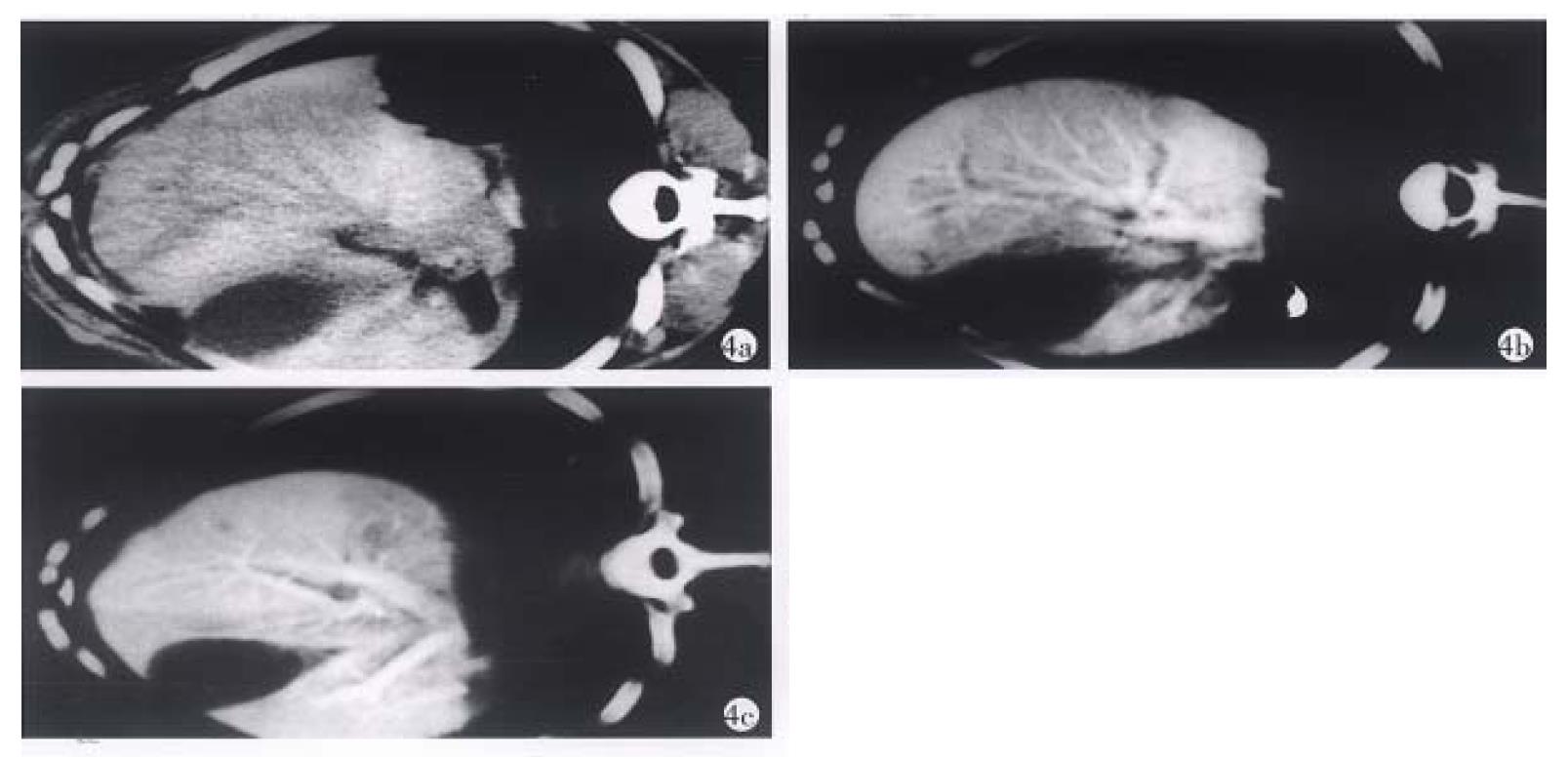
Figure 4 a-c The CT images of the same dog’s liver.
a. plain CT scan; b. CTAP; c. CTSP.
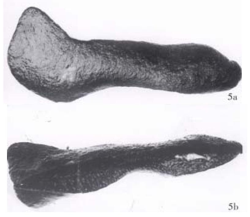
Figure 5 The spleens of the dogs eviscerated 5 d after CTSP.
a. The surface of the spleen is smooth and b. there is nobleeding foci in the longitudinally dissected spleen.
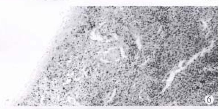
Figure 6 The splenic tissue of one dog killed immediately after CTSP.
HE stain 3.3 × 10.
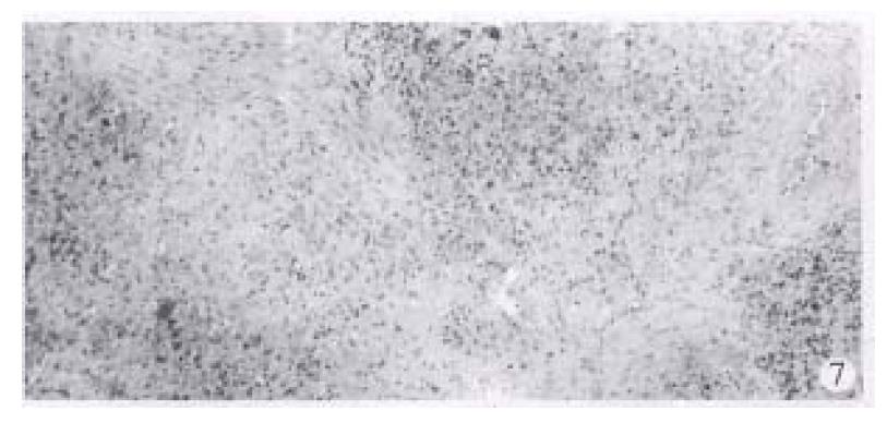
Figure 7 The spleen of a dog killed 10 d after CTSP.
A little proliferation of connecuive tissue could be seen at the point where the catheter entered the spleen. HE stain 3.3 × 10.
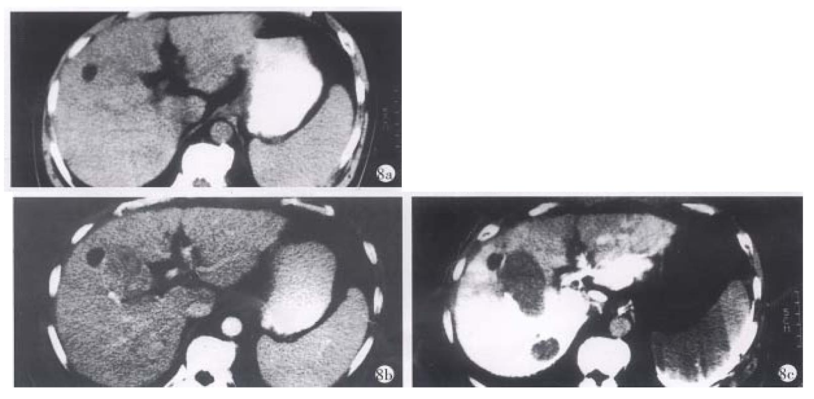
Figure 8 PHC of a 59-year-old male patient.
a. plain CT scan: only a small cyst was seen in the anterior part of the right lobe of the liver. b. enhanced CT: except the cyst, the carcinoma could not be shown clearly. c. CTSP: two carcinomas were clearly shown behind the cyst, and proved to be primary hepatocyte carcinoma.
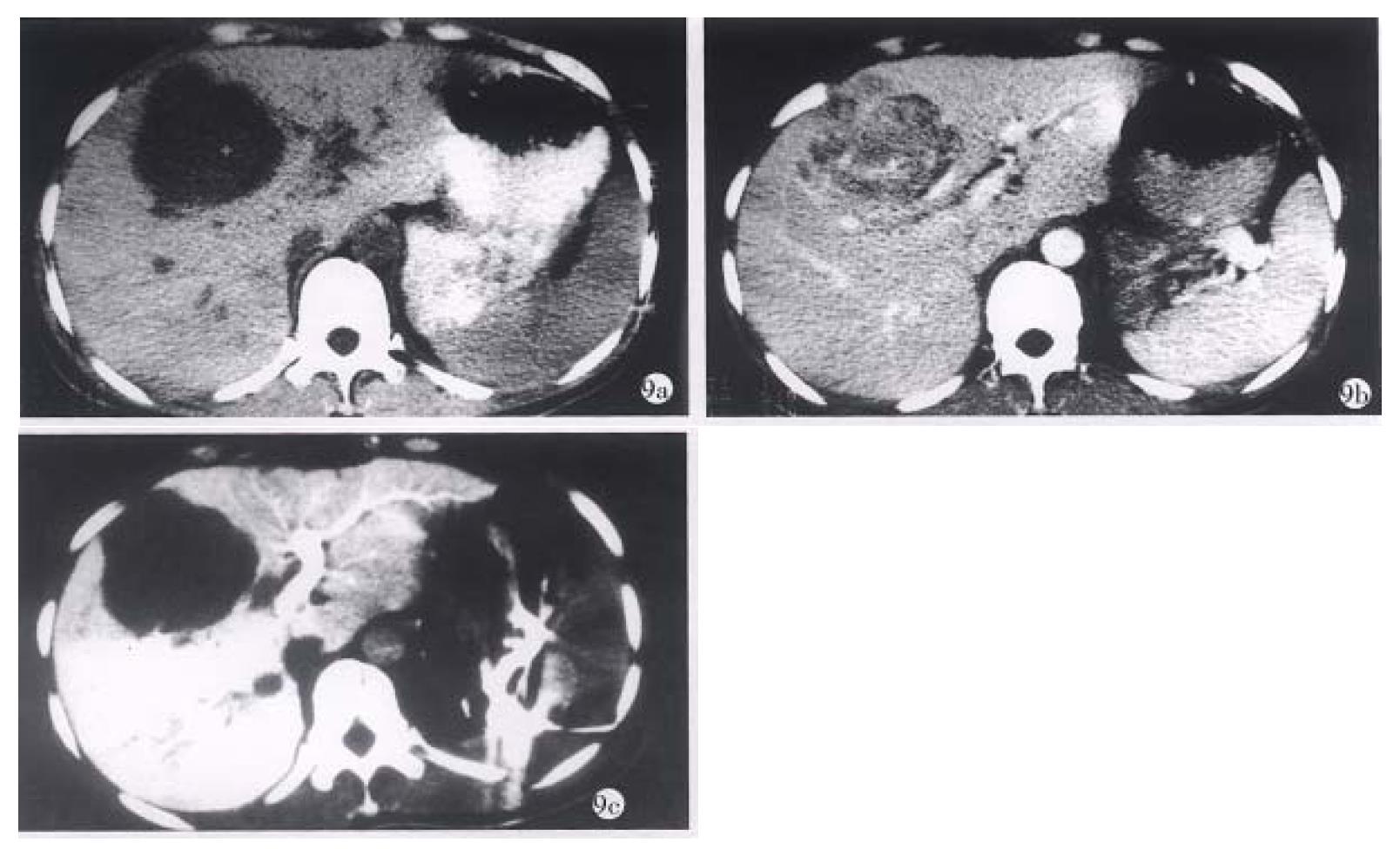
Figure 9 Primary hepatocyte carcinoma of a 30-year-old female patient.
a. plain CT scan; b. enhanced CT; c. CTSP. Though they all could show carcinoma, CTSP not only could show it more clearly than the other two, but also show metastases to the right of the inferior vena cava.
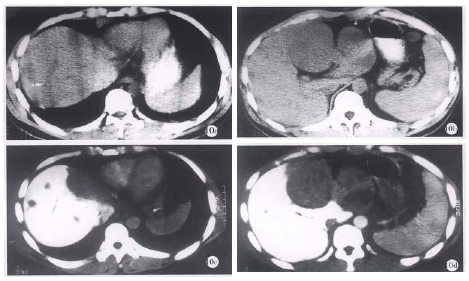
Figure 10 Postoperative recurrence and hepatic metastases of primary hepatocyte carcinoma of a 44-year-old female patient.
a, b, plain CT scans; c, d, CTSP.
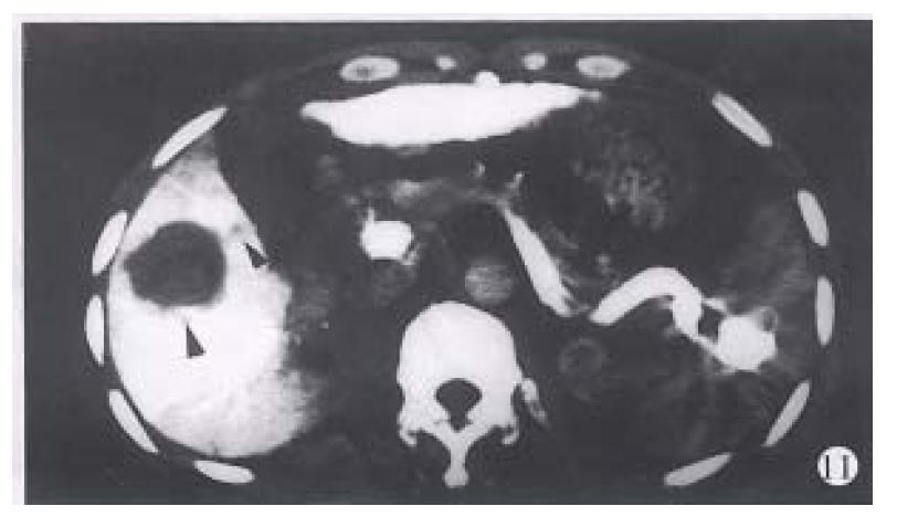
Figure 11 The contrast medium remained in the spleen after CTSP.
The splenic veins show high density. The recurrent cancer in the liver and the child focus in the portal vein (the left posterior arrow) are both shown clearly.
- Citation: Zhang XL, Qiu SJ, Chang RM, Zou CJ. Animal experiments and clinical application of CT during percutaneous splenoportography. World J Gastroenterol 1998; 4(3): 214-218
- URL: https://www.wjgnet.com/1007-9327/full/v4/i3/214.htm
- DOI: https://dx.doi.org/10.3748/wjg.v4.i3.214


