Copyright
©The Author(s) 1998.
World J Gastroenterol. Apr 15, 1998; 4(2): 150-152
Published online Apr 15, 1998. doi: 10.3748/wjg.v4.i2.150
Published online Apr 15, 1998. doi: 10.3748/wjg.v4.i2.150
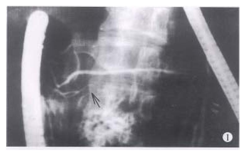
Figure 1 The ERCP manifestations of IPD (↑: communicating branch).
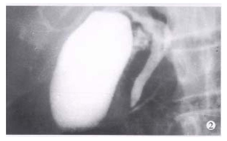
Figure 2 The ventral ductograms of CPD at conventional ERCP.
The duct was short, tapered and arborized into a fine side branches without communicating with the body and tail of the pancreas.
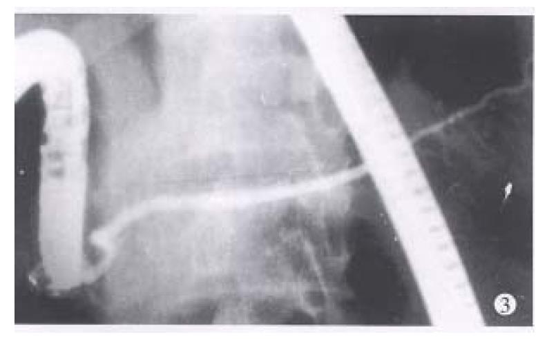
Figure 3 The dorsal ductograms of the above case via the minor papilla cannulation.
The duct was dominant and trended from the pancreatic head to tail.
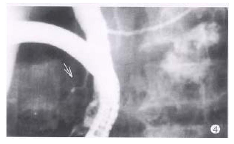
Figure 4 A permanent stenosis of the dorsal ductal terminal was present at the ductograms via the minor papilla cannulation (↑).
- Citation: Lu WF. ERCP and CT diagnosis of pancreas divisum and its relation to etiology of chronic pancreatitis. World J Gastroenterol 1998; 4(2): 150-152
- URL: https://www.wjgnet.com/1007-9327/full/v4/i2/150.htm
- DOI: https://dx.doi.org/10.3748/wjg.v4.i2.150


