Copyright
©The Author(s) 1998.
World J Gastroenterol. Feb 15, 1998; 4(1): 66-69
Published online Feb 15, 1998. doi: 10.3748/wjg.v4.i1.66
Published online Feb 15, 1998. doi: 10.3748/wjg.v4.i1.66
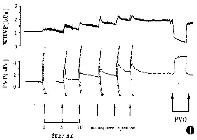
Figure 1 Portal venous pressure (PVP) and wedged hepatic venous pressure (WHVP) recordings obtained from a rat in Group 1 during intraportal microsphere injections and subsequent portal venous occlusion (PVO).
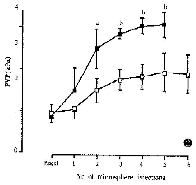
Figure 2 Changes in portal venous pressure (PVP) following intraportal microsphere injections in Group 1(□) and Group 2 (■).
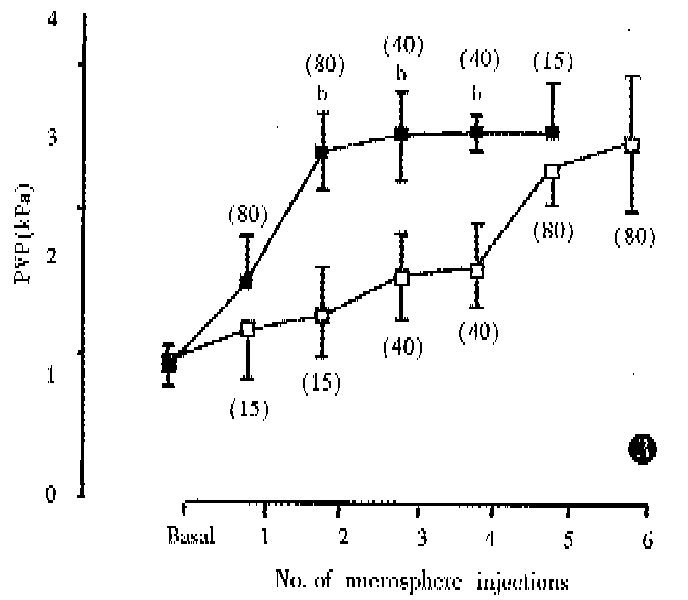
Figure 3 Changes in portal venous pressure (PVP) following intraportal microsphere injections in Group 3(□) and Group 4(■).
Four rats in Group 4 died after the 6th injection and therefore steady PVP recordings could not be obtained after this time. bP < 0.01, Group 4 vs Group 3.
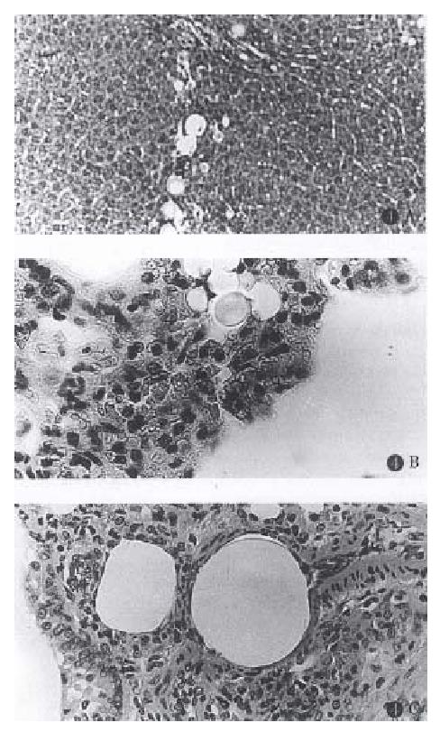
Figure 4 Histological sections showing (A) 15, 40 and 80 μm spheres lodged in liver (Group 3, original magnification × 10), (B) 15 μm spheres lodged in lung (Group 1, original magnification × 40) and (C) 80 μm spheres lodged in lung (Group 2, original magnification × 16).
- Citation: Li XN, Benjamin I, Alexander B. A new rat model of portal hypertension induced by intraportal injection of microspheres. World J Gastroenterol 1998; 4(1): 66-69
- URL: https://www.wjgnet.com/1007-9327/full/v4/i1/66.htm
- DOI: https://dx.doi.org/10.3748/wjg.v4.i1.66


