Copyright
©The Author(s) 1998.
World J Gastroenterol. Feb 15, 1998; 4(1): 33-37
Published online Feb 15, 1998. doi: 10.3748/wjg.v4.i1.33
Published online Feb 15, 1998. doi: 10.3748/wjg.v4.i1.33
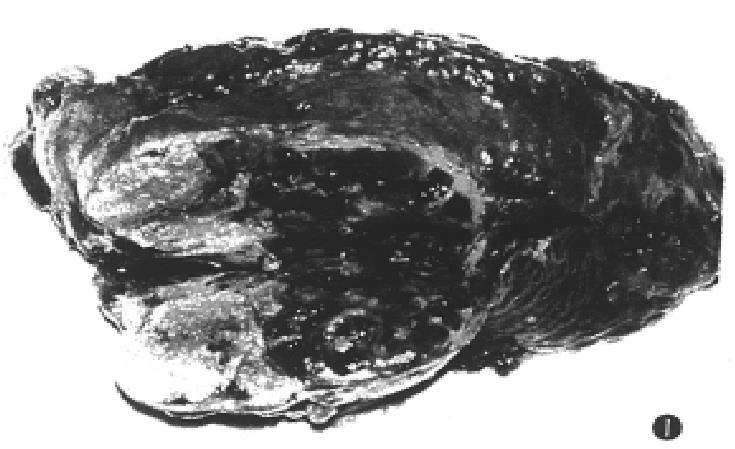
Figure 1 The resected specimen showing massive coagulation necrosis and fibrosis in tumor.
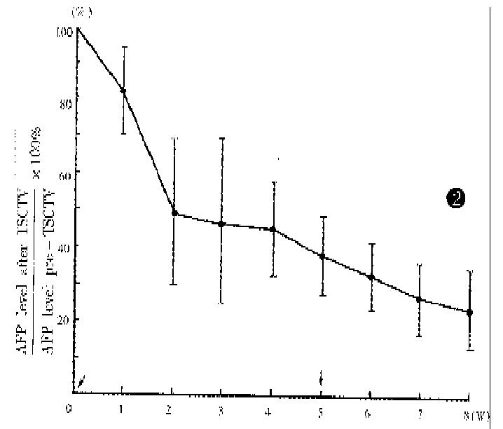
Figure 2 Serum AFP levels in 42 cases (AFP > 400 μmol/L) after TSCTV, assuming that pre-embolization value was 100% (↓TSCTV treatment).
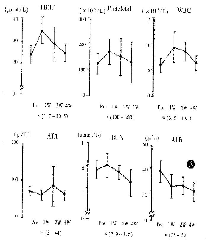
Figure 3 Changes in blood chemical data after first TSCTV (pre: level before TSCTV; W: week; *: normal range).
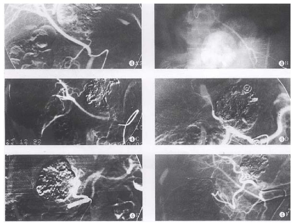
Figure 4 A.
The hepatic right artery derived from superior mesenteric artery after TSCTV was performed. The left hepatic artery derived from gastroduodenal artery and supplying left and lower 1/3 of the tumor, to prevent embolic agents reflux, lipiodol chemoembolization (LCE) was performed. B. The tumor vessels disappeared completely and the lipiodol accumulated within tumor after embolization. C. The lipiodol was obviously washed out in the LCE area after 4 months, TSCTV and LCE were repeated. D. The lipiodol was washed out fourth in the LCE area after 6 months. E. LUF 5 mL and SBMs 50mg were infused through hepatic left artery, the tumor vessels completely disappeared, the LUF accumulated in the LCE area. F. The tumor obviously shrank (PR) and lipiodol accumulated within tumor after 19 months.
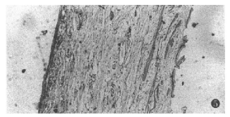
Figure 5 The necrosis of the gallbladded mucosa.
- Citation: Fan J, Ten GJ, He SC, Guo JH, Yang DP, Weng GY. Arterial chemoembolization for hepatocellular carcinoma. World J Gastroenterol 1998; 4(1): 33-37
- URL: https://www.wjgnet.com/1007-9327/full/v4/i1/33.htm
- DOI: https://dx.doi.org/10.3748/wjg.v4.i1.33


