Copyright
©The Author(s) 1998.
World J Gastroenterol. Feb 15, 1998; 4(1): 14-18
Published online Feb 15, 1998. doi: 10.3748/wjg.v4.i1.14
Published online Feb 15, 1998. doi: 10.3748/wjg.v4.i1.14
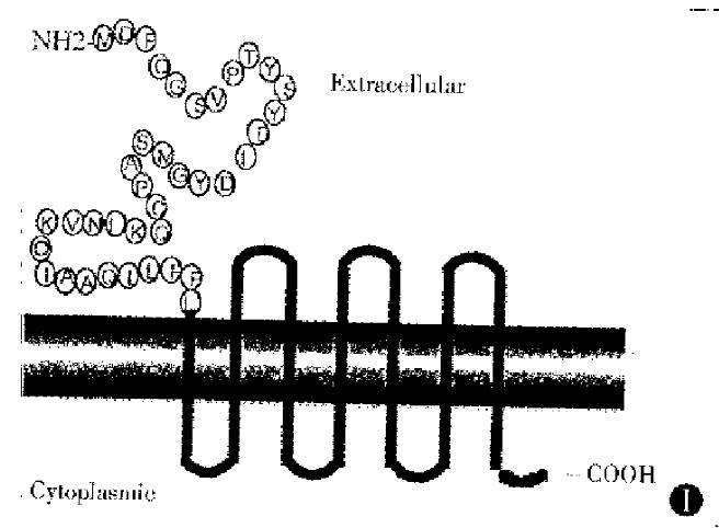
Figure 1 The first extracellular loop amino acid sequence and transmembrane structure of muCCR5.
The amino acid fragment from M to L is located in the first loop of extracellular and it is the binding domain of mouse CCR5 for ligand (MIP-1α, MIP-1β and RANTES).
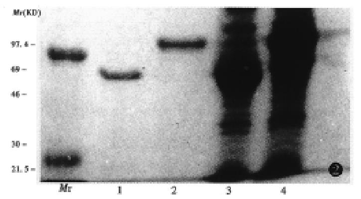
Figure 2 Analysis of recombinant GST-muCCR5-NH2 terminal fusion protein.
Mr: MW marker, 1, 2: products purified by glutathione sepharose, 3, 4: products not purified, 1, 3: expression products induced by IPTG
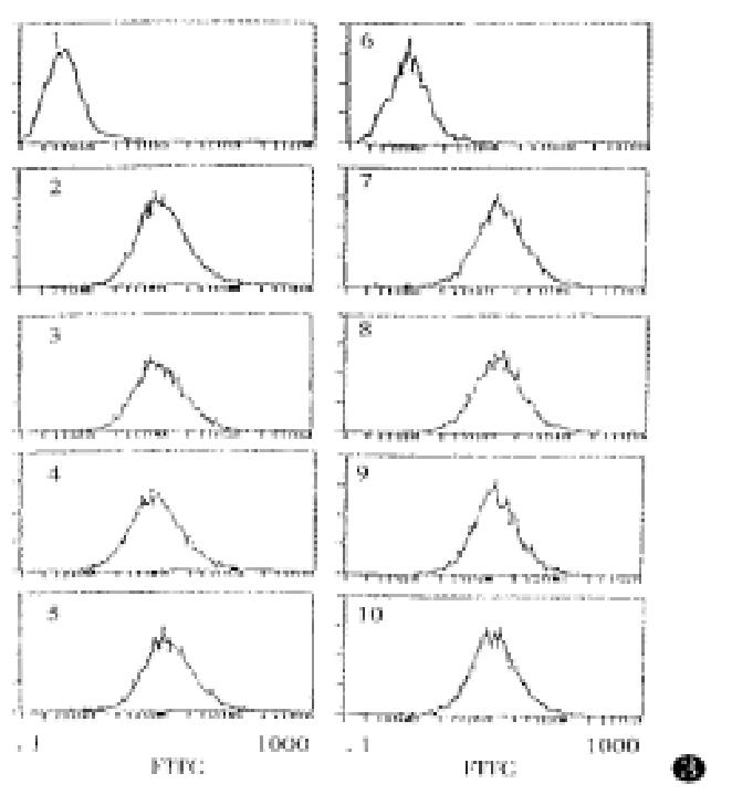
Figure 3 Flow cytometric analysis of specificity of anti-muCCR5 antibody F(ab’)2.
F(ab’)2 volume is 40 mg/L, blocking reagent volume is 100 mg/L. 1. 293 cells; 2. transfected cells of muCCR5. Transfected cells were blocked by: 3. GST; 4. muIL-8; 5. huFusin; 6. GST-muCCR5-NH2; 7. muCCR1; 8. huCCR2; 9. huCCR3 and 10. huCCR4.
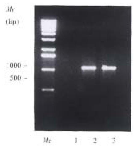
Figure 4 RT-PCR analysis of DTH reactions.
muCCR5 specific primers were used to amplify the cDNA from spleen cells. Lane. 1. BALB/C moase fulminant hepafifis induced by P. acnes, Lane 2.24 h result after 1% picryl chloride stimulation. Lane 3.48 h result after 1% picryl chloride stimulation.
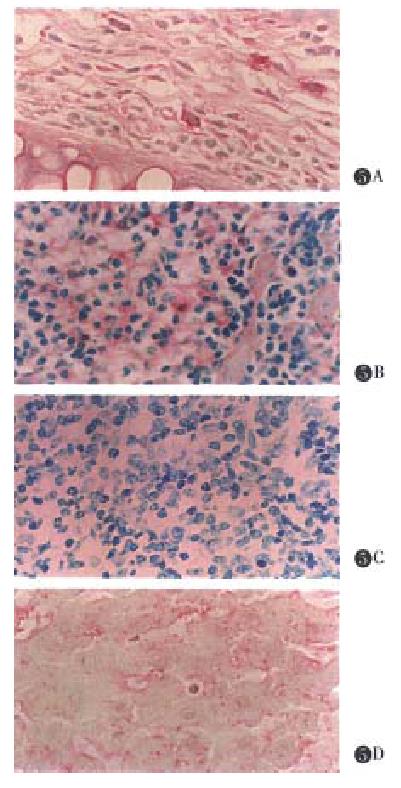
Figure 5 Immunohistochemical analysis of DTH reaction.
A.Ear: Most of the macrophages in subepidermal interstitial tissue and in the local capillary were obviously positive in their cytoplasm for the staining. S. Spleen: There were plenty of reticular-macrophage in red medulla. A few slightly positive cells, however, could be seen scattered there. C. The negative result was used by anti-GST F(ab’)2. D. Liver: The cytoplasm of Kupffer cells were slightly stained by the dyes.
- Citation: Guo BY, Zhang SY, Mukaida N, Harada A, Kuno K, Wang JB, Sun SH, Matshshima K. CCR5 gene expression in fulminant hepatitis and DTH in mice. World J Gastroenterol 1998; 4(1): 14-18
- URL: https://www.wjgnet.com/1007-9327/full/v4/i1/14.htm
- DOI: https://dx.doi.org/10.3748/wjg.v4.i1.14


