Copyright
©The Author(s) 2025.
World J Gastroenterol. Feb 7, 2025; 31(5): 98732
Published online Feb 7, 2025. doi: 10.3748/wjg.v31.i5.98732
Published online Feb 7, 2025. doi: 10.3748/wjg.v31.i5.98732
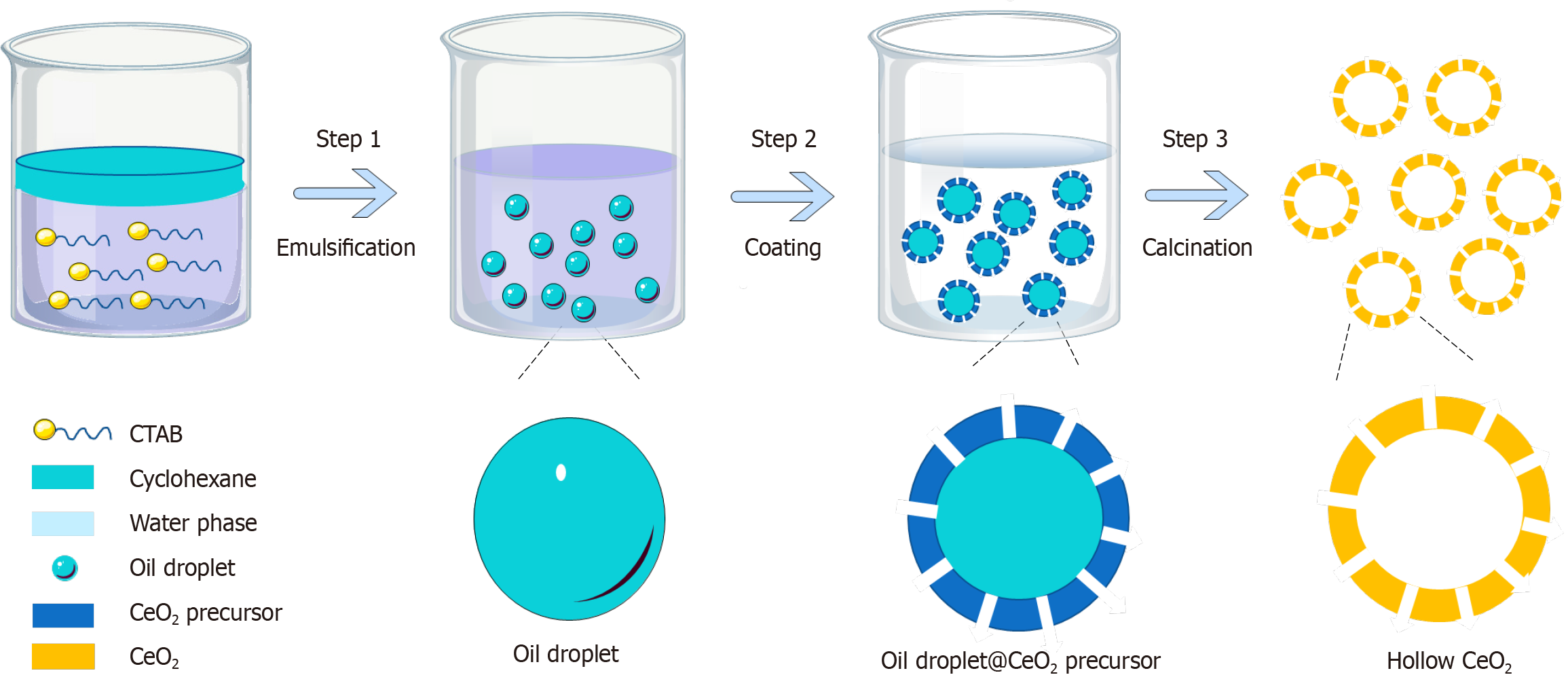
Figure 1
Hollow cerium nanoparticles was synthesized by the one-step method.
Figure 2 Characterization of hollow cerium nanoparticles.
A: Transmission electron microscopy image of hollow cerium (H-CeO2) nanoparticles; B: Scanning electron microscope image of H-CeO2; C: Dynamic light scattering image of H-CeO2.
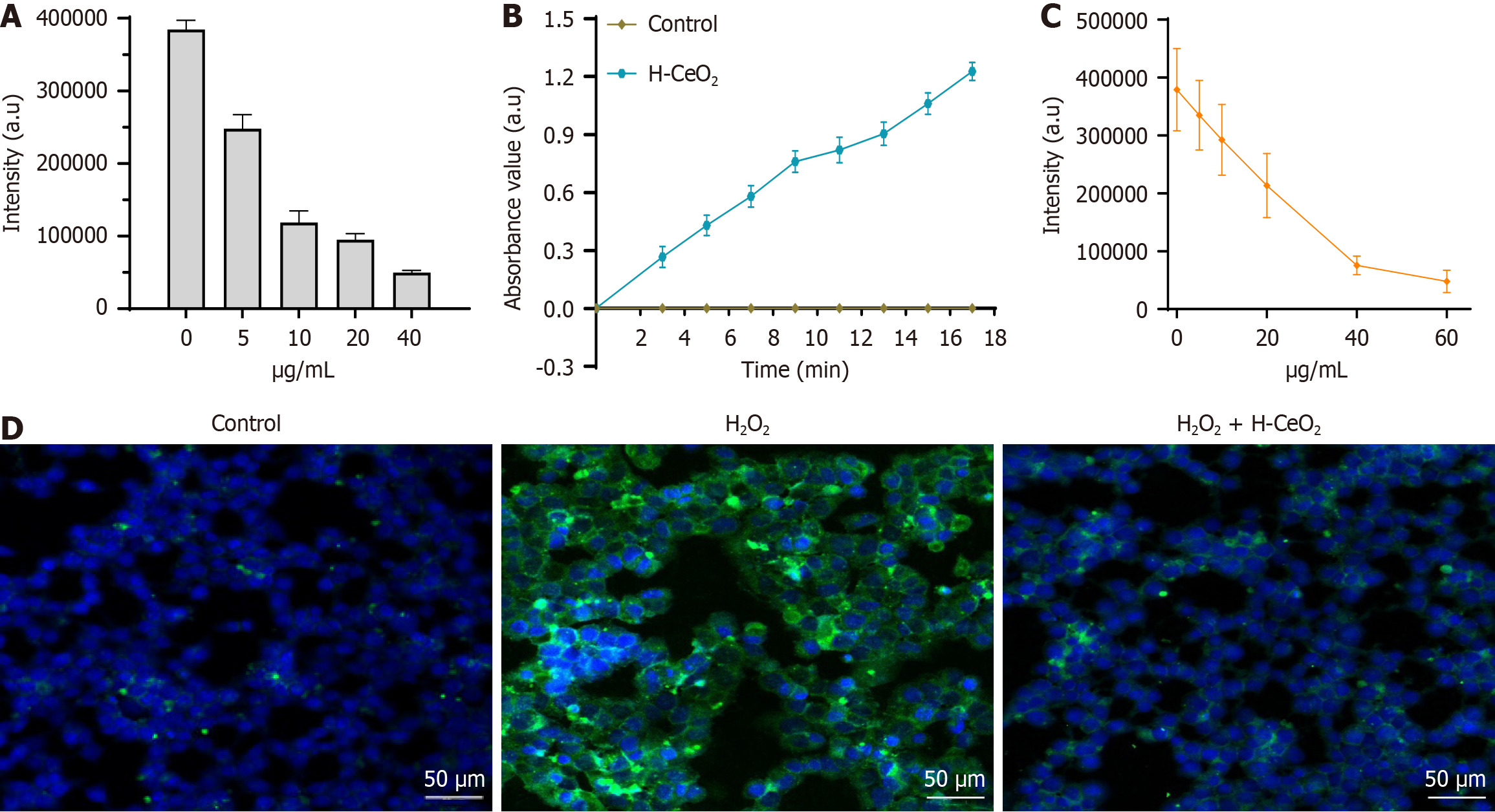
Figure 3 Hollow cerium nanoparticles exhibits good elimination of ∙OH, ∙OOH and reactive oxygen species in vitro.
A: The effect of hollow cerium (H-CeO2) nanoparticles on the formation of ∙OH in the TiO2/UV system; B: Peroxidase activity of H-CeO2; C: Effects of H-CeO2 on ∙OOH produced by the xanthine/xanthine oxidase system; D: Dichlorofluorescein Diacetate probe to detect the ability of H-CeO2 to scavenge reactive oxygen species in HCT116 cells. H-CeO2: Hollow cerium.
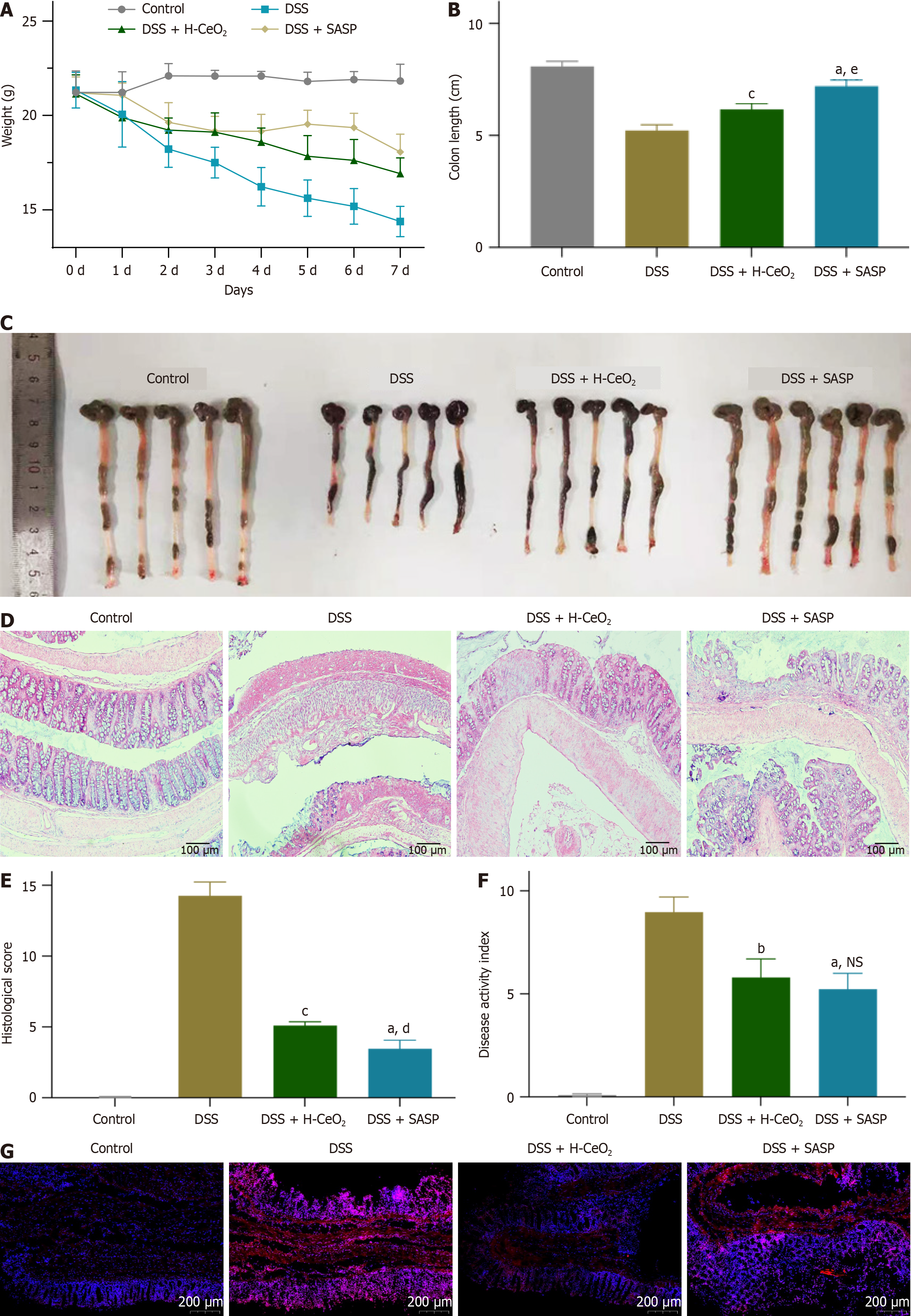
Figure 4 Hollow cerium inhibits occurrence and development of colitis in mice by scavenging reactive oxygen species.
A-C: Hollow cerium nanoparticles (H-CeO2) treatment reduced the changes in weight (A) and colon length (B and C) of dextran sulfate sodium (DSS)-induced colitis mice; D and E: Hematoxylin and eosin detected the effect of H-CeO2 treatment on the histopathology of the colon in DSS-induced mice; F: H-CeO2 treatment reduces disease activity index values in DSS-induced mice; G: H-CeO2 treatment reduced reactive oxygen species levels in DSS-induced mouse tissues. Cell nuclei stained with 4’,6-diamidino-2-phenylindole (blue), and intracellular reactive oxygen species levels were quantitated in dihydroethidium (red). aP < 0.001 vs control group; bP < 0.01 vs dextran sulfate sodium group; cP < 0.001 vs dextran sulfate sodium group; dP < 0.01 vs dextran sulfate sodium + hollow cerium nanoparticles group; eP < 0.001 vs dextran sulfate sodium + hollow cerium nanoparticles group; NS indicates statistical difference. DSS: Dextran sulfate sodium; H-CeO2: Hollow cerium; SASP: Sulfasalazine; NS: Not significant.
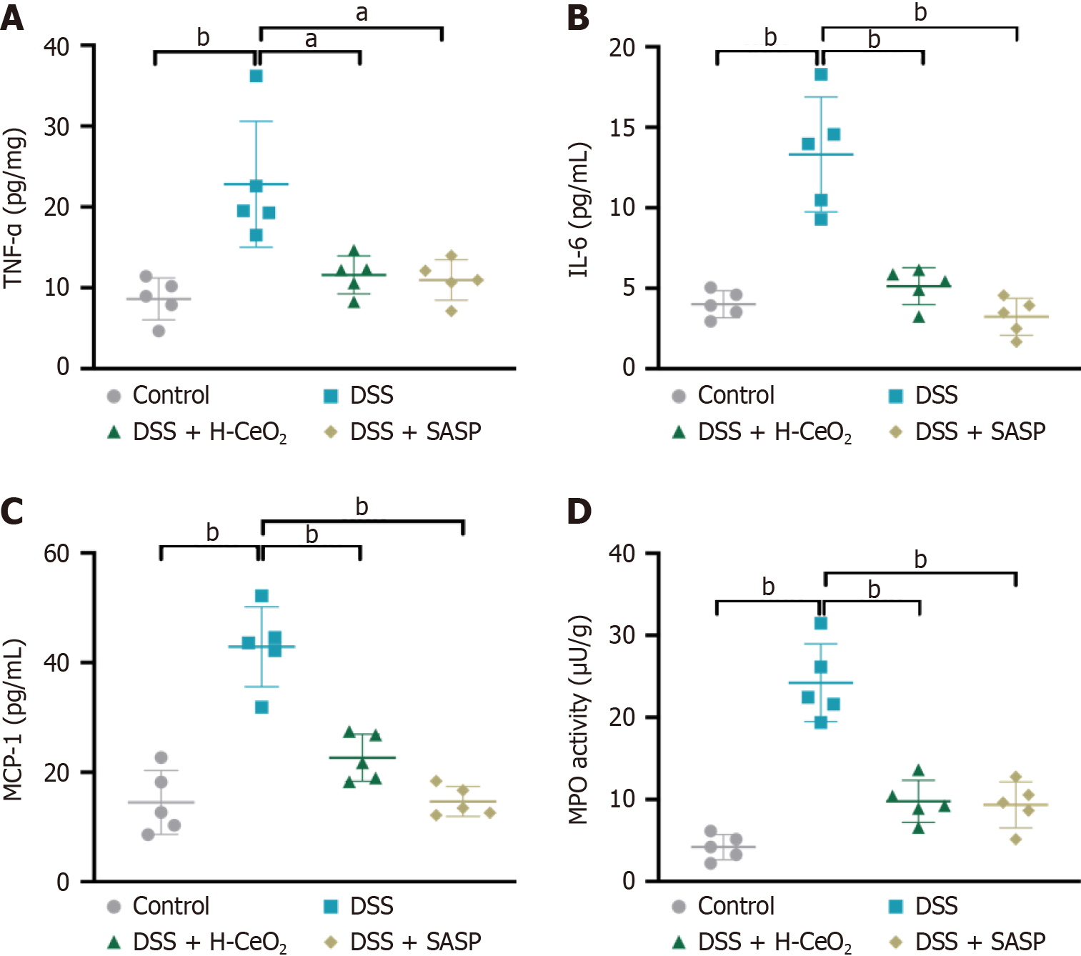
Figure 5 Hollow cerium inhibits inflammatory factors.
A-C: The expression levels of pro-inflammatory factors tumor necrosis factor-α (A), interleukin-6 (B), and monocyte chemoattractantprotein-1 (C) in serum were detected by enzyme-linked immunosorbent assay; D: Myeloperoxidase activity in serum. aP < 0.01; bP < 0.001. DSS: Dextran sulfate sodium; H-CeO2: Hollow cerium; SASP: Sulfasalazine; TNF: Tumor necrosis factor; IL: Interleukin; MCP: Monocyte chemoattractan
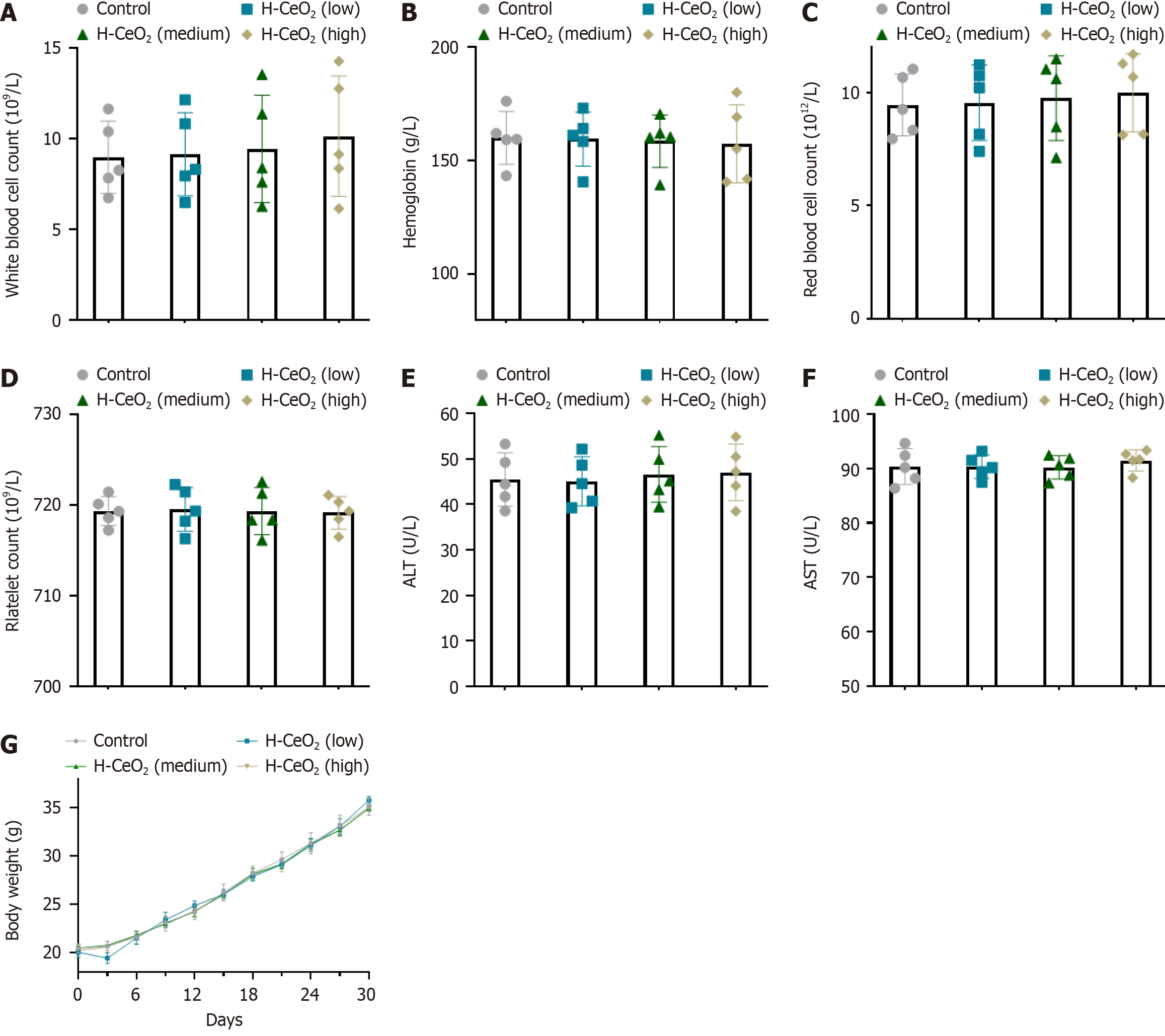
Figure 6 Hollow cerium has good biological safety.
A-F: The effect of hollow cerium nanoparticles on the levels of blood routine (A-D) and liver function indexes alanine aminotransferase (E) and aspartate aminotransferase (F) in the serum of healthy mice; G: Hollow cerium nanoparticles did not affect the bodyweight of healthy mice. H-CeO2: Hollow cerium; AST: Aspartate aminotransferase; ALT: Alanine aminotransferase.
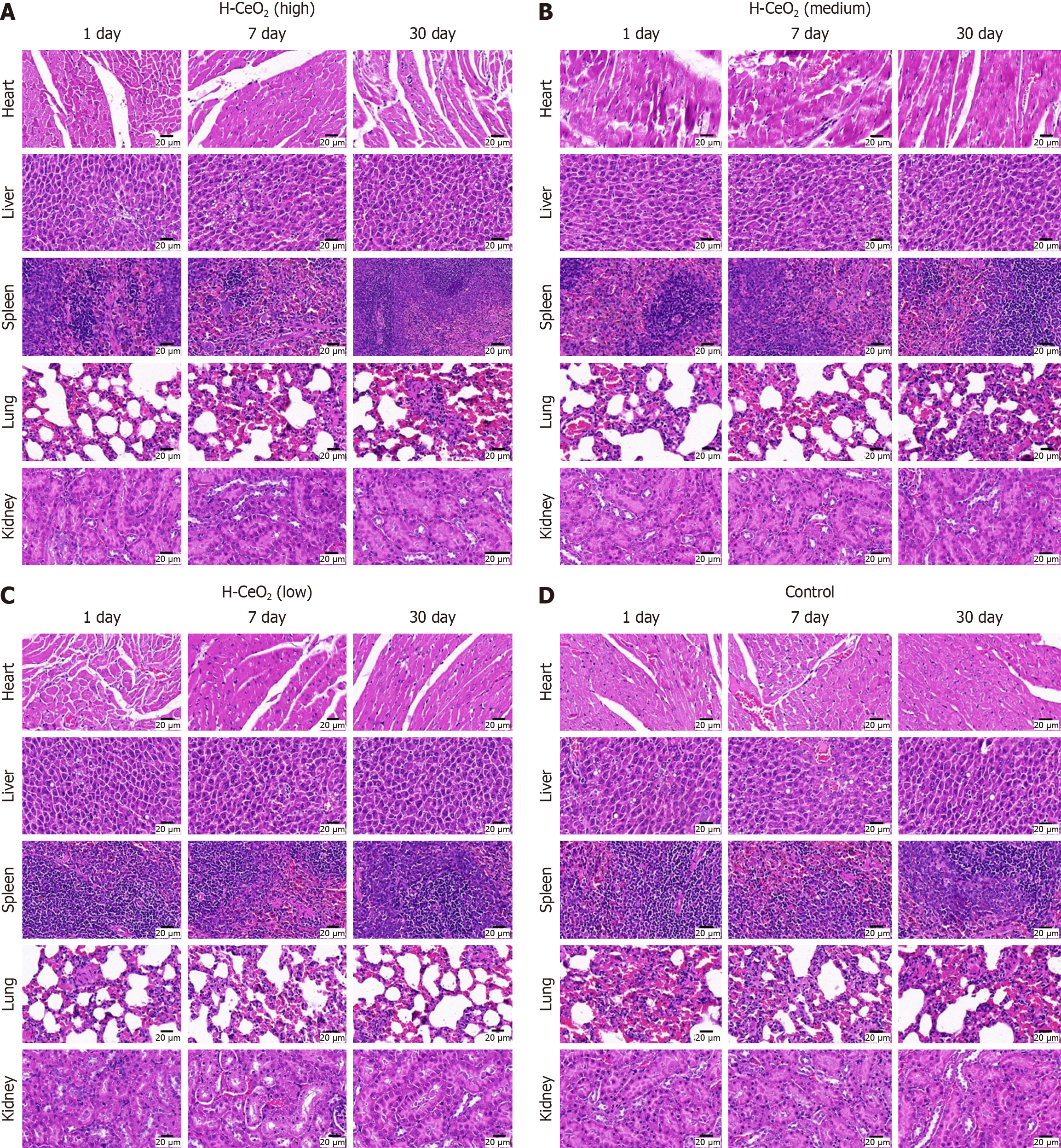
Figure 7 Hematoxylin and eosin detected the effect of different doses of hollow cerium nanoparticles administration on the main organs of healthy mice.
A-D: Hematoxylin and eosin staining of the main organs from mice receiving 2 mg/mL (A), 1 mg/mL (B), 0.01 mg/mL (C), and saline (D) injection. H-CeO2: Hollow cerium.
- Citation: Mi L, Zhang K, Ma JX, Yao JF, Tong YL, Bao ZJ. Hollow cerium nanoparticles synthesized by one-step method for multienzyme activity to reduce colitis in mice. World J Gastroenterol 2025; 31(5): 98732
- URL: https://www.wjgnet.com/1007-9327/full/v31/i5/98732.htm
- DOI: https://dx.doi.org/10.3748/wjg.v31.i5.98732


